Product Information
Exosomes are secreted nanovesicles with diameters ranging from 30 to 150 nm and produced in the endosomal compartment of most eukaryotic cells. They are present in all body fluids under both normal and pathophysiological conditions. They have important roles in mediating intercellular communication by serving as carriers of different biomolecules, including proteins, RNAs, and lipids from one cell to another. These unique properties of exosomes provide opportunities for innovations in diagnosis and treatments.
Many samples, such as cell culture media, saliva, and urine, contain exosomes at low concentrations. To maximize efficiency, GeneCopoeia provides ExoSure™ Exosome Isolation Kit, which combine precipitation and size exclusion chromatography (SEC) techniques, generating high purity and high yields of exosomes.
Advantages
- High yield. Up to 5 x 10¹⁰ of purified exosomes per column, depending on cell types and cell culture conditions.
- High quality. Preserving extracellular vesicles (EVs) original shape and biological function based on size exclusion chromatography (SEC) method.
- Fast and cost-effective. Requires less than 20-minutes of hands-on time, including a convenient column-based clean-up.
- Widely used. Available for electron microscopy analysis, Nanoparticle Tracking Analysis (NTA) analysis, nucleic acid and protein analysis, in vitro and in vivo experiments, etc.
Product Details
Performance
Size distribution of ExoSure™-isolated exosomes measured by NTA shows high yield of exosomes isolation. (cell supernatant)
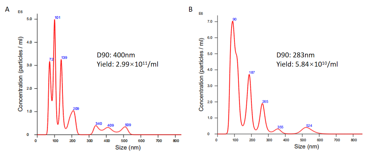
Figure 1. NTA data of ExoSure™-isolated exosomes from HEK293T cells. Size and particle concentration distribution plot of exosomes from HEK293T cells (peak at 101 nm). D90 value is 400nm using ExoSure™ Solution (Cat. No. EP001) (A) and (peak at 90 nm), D90 value is 283nm using ExoSure™ Exosome Isolation Kit (Cat. No. EP002) (B).
Size distribution of ExoSure™-isolated exosomes measured by NTA shows high yield of exosomes isolation. (serum)
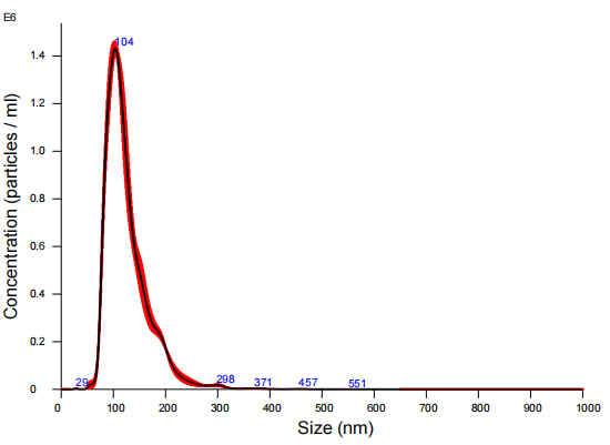
Figure 2. NTA data of ExoSure™-isolated serum exosomes. Purified serum exosomes (peak at 104 nm), D90 value is 180nm using ExoSure™ Exosome Isolation Kit (Cat. No. EP002).
CD63 exosome biomarker western blot assay.
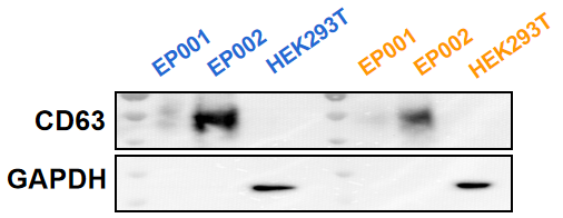
Figure 3. CD63 exosome biomarker western blot. Exosome from HEK293T cultured media was isolated by ExoSure™ Exosome Isolation Kit (Cat.No. EP001 and EP002). Both of exosome and HEK293T cell lysates with 10μg(in blue)/5μg(in orange) were detected by anti-CD63 and anti-GAPDH antibody.
Performance comparison of ExoSure™ with competitor’s kit by luciferase assays.
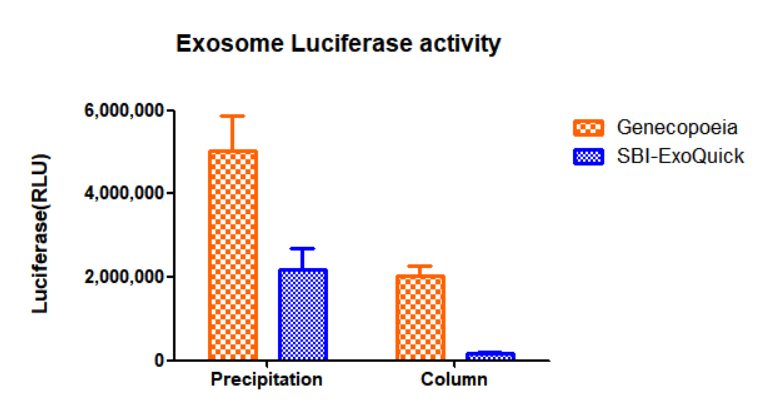
Figure 4. Luciferase activity assays. Exosomes were isolated from HEK293T cells transfected with Luciferase-CD63 plasmids using GeneCopoeia ExoSure™ Exosome Isolation Kit and System Biosciences (SBI) ExoQuick-TC® ULTRA EV Isolation Kit. The Luciferase activity of exosomes were tested using Luciferase Assay Kit (GeneCopoeia, Cat. No. LF061).
FAQs
Frequently Asked Questions: Exosome Isolation
Answer: The ExoSure™ Exosome Isolation Kit technology combines precipitation and size exclusion chromatography (SEC) columns. SEC is a method to separate particles in solution based on their size. The chromatography column is packed with stable polymeric beads, creating a porous matrix. When the solution containing exosomes is added to the SEC column, the smaller particles will be trapped in the pores, while larger molecules do not enter the pores and elute first. As a result, different elution fractions will contain molecules of different sizes; first the large particles, followed by the smaller particles.


 Powered by Bioz