GeneCopoeia’s OmicsArray™ antigen microarrays are high-throughput protein microarrays (also known as “protein arrays” or “protein chips”) that are powerful tools for many applications, including autoantibody profiling, biomarker detection, pathogen-specific antibody identification, and more. Antigen microarrays enable the detection of hundreds to thousands of antibodies or other biomarkers present in multiple samples in parallel.
Each array contains superior-quality purified antigens printed onto nitrocellulose filters, which are adhered to glass slides. Antigens known to be associated with specific diseases or pathogens are chosen based on a thorough review of peer-reviewed publications.
In addition to premade arrays, arrays containing customized sets of antigens are available, as well as array processing kits, array profiling services, and data analysis.
Advantages of OmicsArray™ Antigen Microarrays
Largest collection of pre-made whole-protein antigen microarrays on the market.
Largest number of whole-protein antigens specifically focused on autoimmune disease research.
Best combination of number of antigens per array (up to 120) with number of samples that can be processed per slide (up to 15).
GeneCopoeia offers custom antigen microarray services in the following areas:
Custom array printing. GeneCopoeia will create custom antigen microarrays built to your specifications.
Sample processing. Send us your blood, plasma, tissue, or other biological sample and we will prepare it for processing and incubation with any of our premade antigen microarrays or custom-built antigen microarrays for autoantibody profiling and other applications. For information on sample types to submit, consult the FAQ
Data analysis. Once samples are processed and incubated with an antigen microarray, we will analyze the raw data. The standard analysis service includes: 1) An Excel file of the Net Signal Intensity (NSI) for each antigen on the array, normalized to internal controls; and 2) a heat map
Additional analysis services, including proteomic analysis, pathway analysis, and more, are also available.
To inquire about custom antigen microarray products and services,please fill out our custom quote request form.
GeneCopoeia’s OmicsArray™ antigen microarrays contain up to 120 purified antigens spotted onto nitrocellulose filters, which are adhered to glass slides. In addition, 8 spots are included for normalization. Each slide carries 16 identical arrays, and so can be used to process up to 15 samples simultaneously as well as a negative control. As little as 1 μL serum or 50 μL of other bio fluids is needed for each sample.
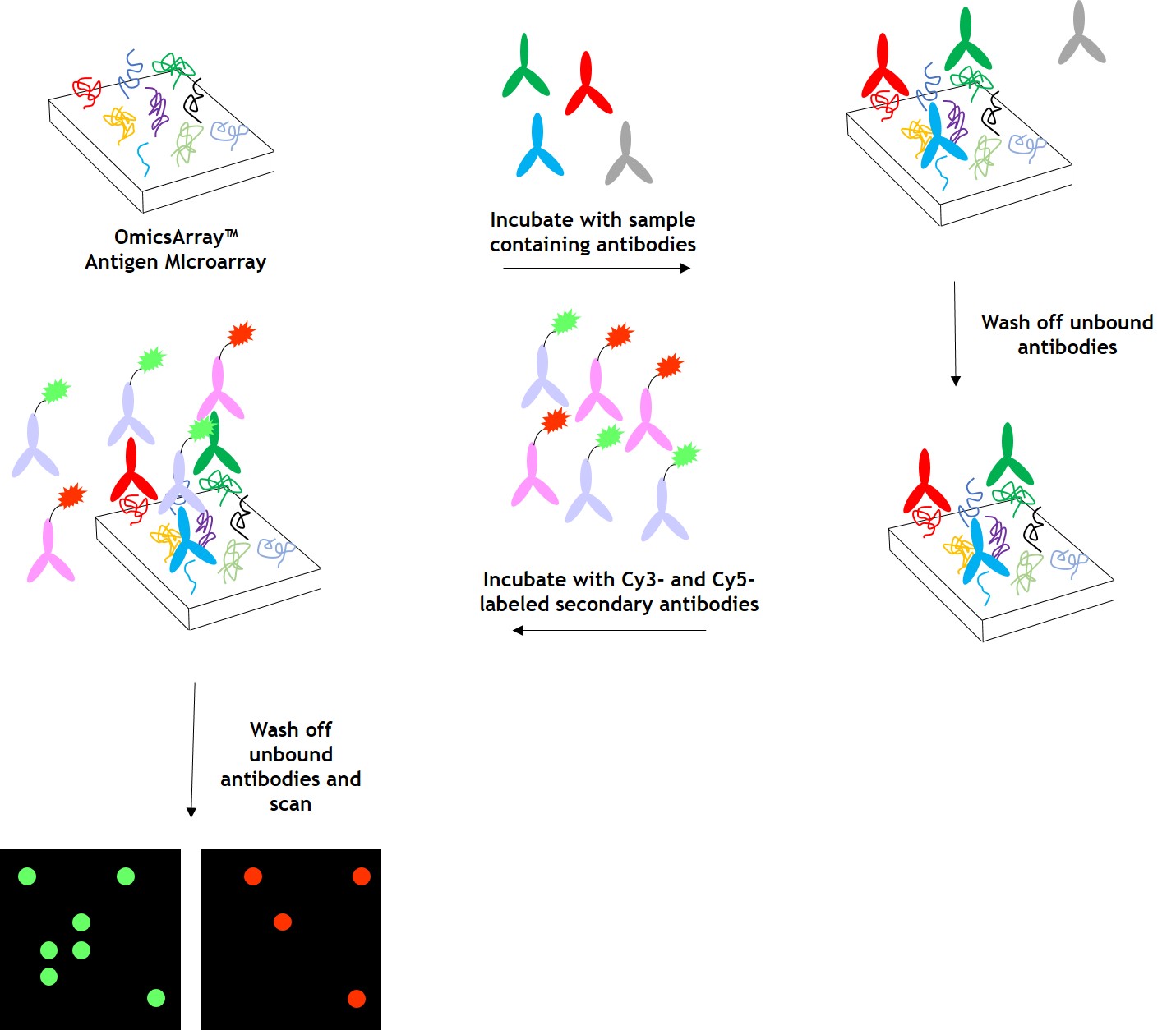
As shown in Figure 1, arrays are incubated with patient samples, and any autoantibodies in the samples bind to their cognate antigens on the array. The arrays are washed to remove unbound autoantibodies and other proteins, then co-incubated with Cy3- and Cy5-labeled secondary antibodies. The dual labeling strategy is intended to distinguish between immunoglobulin (Ig) subtypes present within samples. For example, a Cy3-labeled anti-IgG secondary antibody is used to detect IgG autoantibodies, and a Cy5-labeled anti-IgM secondary antibody is used to detect IgM autoantibodies. Fluorophore-labeled secondary antibodies are available for detecting IgA, IgD, IgE, IgG and IgM immunoglobulins, as well as IgG subclasses IgG1, IgG2, IgG3, and IgG4.
After washing to remove unbound secondary antibodies, signals are detected using a microarray scanner (e.g., GenePix® 4000B, InnoScan 710, or equivalents). The raw data is then analyzed using GenePix® Pro 7.0 or Mapix software.
Figure 1. Workflow for autoantibody profiling in samples using GeneCopoeia’s OmicsArray™ antigen microarrays.
GeneCopoeia provides data analysis as part of its custom autoantibody profiling services. After array scanning, raw data are collected and analyzed using GenePix® 7.0 software. The standard service includes data normalization to include fold changes of, for example, disease vs. healthy control, and a heat map showing each antigen ranked by level of fold change. Our service undergoes the following steps:
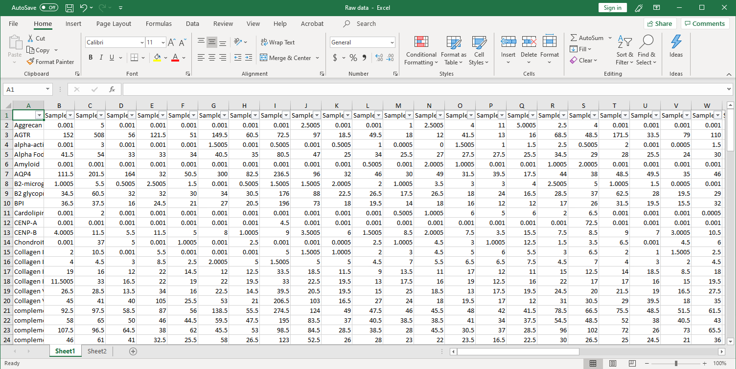
1. Collection of raw data. After scanning, raw signal intensities are collected in an Excel file, as shown below.
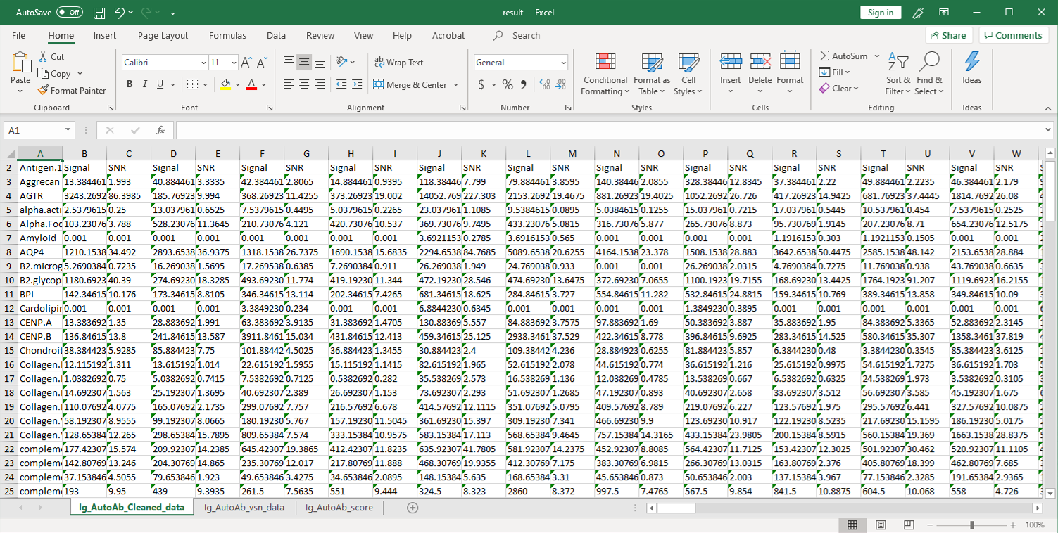
2. Next, the raw data are normalized to controls on the array, and the Net Signal Intensity (NSI) values, as well as the signal-to-noise ratios (SNRs) are tabulated in an Excel file,
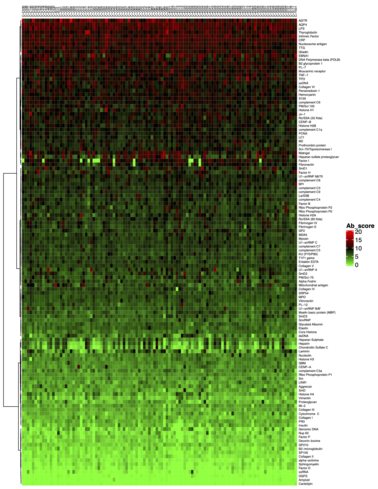
3. The final step of the standard analysis package is to determine the “Antibody score” which is the relative enrichment of a given antibody in a sample. The antibody score is determined numerically and displayed in a heat map. A higher score (shifted toward red on the heat map) suggests a stronger antibody-antigen interaction, as shown in the following example:
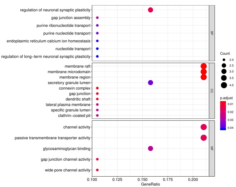
4. In addition to the standard data analysis package, customers can also choose custom data analysis services. One such service is to classify positive antigens on the array using Gene Ontology (GO) analysis, which classifies genes and proteins based on known biological functions. An example of the readout from GO analysis for molecular function is shown below:
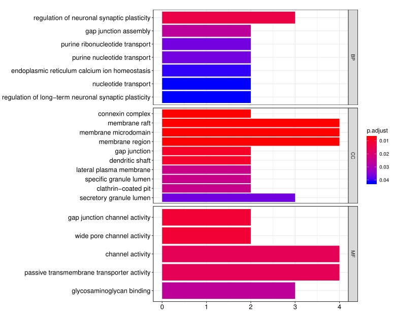
Further, GO analysis for biological processes is shown in the following example:
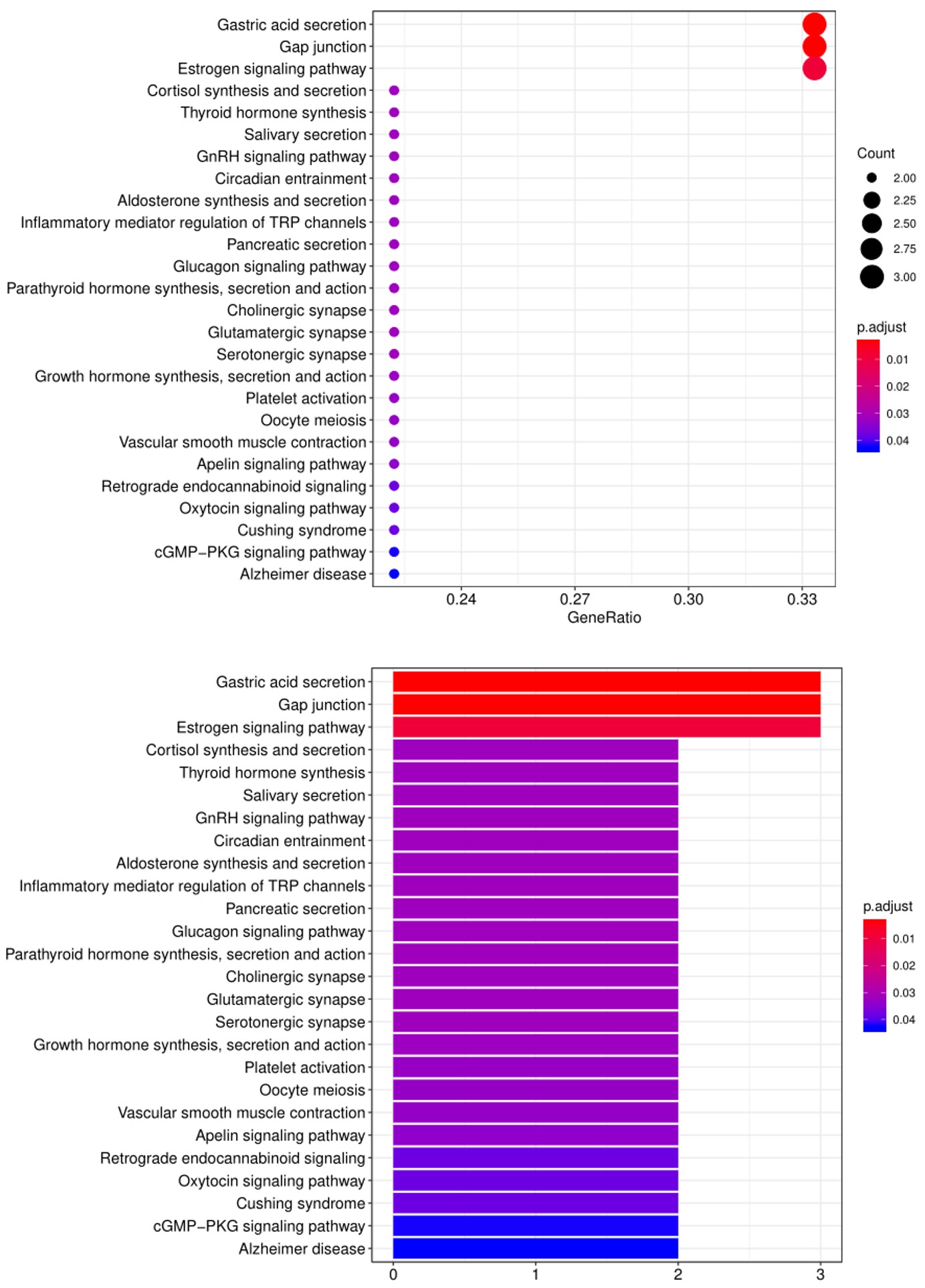
5. Another widely-used tool for bioinformatic analysis is KEGG (Kyoto Encyclopedia of Genes and Genomes) Pathway Analysis, which we use to group positive antibody-antigen interactions based on defined biological pathways, as displayed in the example below:
Answer: You can send us virtually any biological sample, including whole blood, serum, plasma, interstitial fluid, semen, urine, and saliva. To learn more, please contact us at inquiry@genecopoeia.com.
Answer: a. Each slide carries 16 identical arrays. One of these is reserved for a PBS control, while the remaining 15 are used for incubation of each sample. Therefore, the total number of samples sent by the customer is preferably a multiple of 15. However, if you have fewer than 15 samples, we will need to charge you for the full cost of an entire slide of 15 samples. b. Theoretically, each of the different test groups requires a minimum of 3 samples to meet statistical reproducibility requirements. On this basis, the greater the number of samples, the higher the reproducibility of the results in the test group. In order to ensure the reliability of the results, the sample size of different test groups is recommended to be ≥3.
Answer: The antigen microarray uses fluorescence detection, and its sensitivity is higher and more stable than ELISA (colorimetry) and Western (chemiluminescence). The technical repetition correlation coefficient R2 between the arrays can reach 0.9 or more. The dynamic detection range is 1-65000. The larger the dynamic range, the more layers of signals that can be detected.
Answer: Please fully communicate with our Technical Support staff before the experiment to explain the purpose of the experiment and the sample status, to determine whether we can meet your needs and determine the experimental plan.
Answer: The antigens on the antigen array are all derived from self-antigens reported in the literature. For each of the different types of antigenic arrays, we can provide you with all the names of the antigens upon reques Please do not disclose this information to third parties.
Answer: If you need a custom antigen microarray, we need to fully communicate with you about the following issues: a. The intellectual property of the antigen (whether from a published paper or a patent, etc.). We can only provide customized detection of antigens that do not involve patent protection; b. Basic information about the antigen (whether it is whole protein or peptide, molecular weight, domain, whether it is a membrane protein, etc.); c. Whether you can provide an antigen that meets the requirements of a custom array; d. If we need to provide the antigen to be tested, the corresponding cost and experimental time frame need to be accounted for separately.
Answer: a. This difference may be related to the detection method. The protein used on the array is a non-denatured protein, which is different from western blot. The hybrid system of the chip is an antigen-trapping antibody, and the western blot antigen is slightly different from the antibody after interacting with the antigen; b. Please provide a graph of the results of western blot verification, so that we can better analyze the image and data of your verified indicators on the array.
Title: Antigen Protein Microarrays For Effective Antibody Profiling.
Presented Wednesday, September 22, 2021
GeneCopoeia’s OmicsArray™ antigen microarrays are protein microarrays engineered for several powerful applications, including autoantibody detection, biomarker profiling, and characterization of protein-molecule interactions. Antigen microarrays enable parallel detection of antibodies or other biomarkers present in patient samples for diseases including rheumatoid arthritis, muscular dystrophy, systemic lupus erythematosus, and type 1 diabetes. In this webinar, we will describe the powerful application and advantages of using GeneCopoeia’s OmicsArray™ antigen microarrays for autoantibody detection, cancer immune checkpoint inhibition autoantibody profiling, and SARS-CoV-2 serological analysis.
Please enter your email address, then watch the recorded webinar
Title: OmicsArray™ Antigen Microarrays for high-throughput Autoantibody Screening for Autoimmune and Immune-associated Disorders.
Dr. Quanzhen Li from GeneCopoeia throws light on OmicsArray™ Antigen Microarrays for high-throughput Autoantibody Screening for Autoimmune and Immune-associated Disorders through an informative webinar hosted by GeneCopoeia.
SARS-CoV-2 is a betacoronavirus that is the cause of the COVID-19 global pandemic that began in 2019. SARS-CoV-2 infection of humans typically stimulates a strong humoral immunity response, leading to the production of both neutralizing and binding antibodies.
GeneCopoeia provides COVID-19-related antigen microarrays for two purposes: 1) Association of autoimmunity against cytokines with SARS-CoV-2 infection (Coronavirus Associated Autoimunity microarray, catalog # PA012); and 2) Detection of antibodies against SARS-CoV-2 (including variants of concern) and other pathogens (SARS-CoV-2 Coronavirus Variant Proteins microarray, catalog # PA013).
In addition to these predesigned arrays, arrays containing customized sets of proteins are available, as well as array profiling services and data analysis.
Predesigned arraysPremade antigen microarrays GeneCopoeia’s OmicsArray™ COVID-19 Coronavirus Antigen Microarrays are premade protein microarrays. Please refer to the table below for more information.
In addition to premade antigen microarrays, GeneCopoeia offers custom antigen microarray services in the following areas:
Custom printing and spotting. GeneCopoeia will create custom antigen microarrays built to your specifications.
Sample processing. Send us your blood, plasma, tissue, or other biological sample and we will prepare it for processing and incubation with any of our premade antigen microarrays or custom-built antigen microarrays
Data analysis. Once samples are processed and incubated with an antigen microarray, we will analyze the raw data. The standard analysis service includes: 1) An Excel file of the Net Signal Intensity (NSI) for each antigen on the array, normalized to internal controls; and 2) a heat map
Additional analysis services, including proteomic analysis, pathway analysis, and more, are also available.
To inquire about custom antigen microarray services, please contact us at inquiry@genecopoeia.com.
GeneCopoeia’s OmicsArray™ antigen microarrays contain up to 120 purified proteins spotted onto nitrocellulose filters, which are adhered to glass slides. In addition, 8 spots are included for normalization. Each slide carries 16 identical arrays, and so can be used to process up to 15 samples simultaneously as well as a negative control. As little as 1 ul serum or 50 ul of other bio fluids are needed for each sample.
As shown in Figure 1, arrays are incubated with patient samples, and any antibodies in the samples bind to their cognate antigens on the array. The arrays are washed to remove unbound antibodies and other proteins, then co-incubated with Cy3- and Cy5-labeled secondary antibodies. The dual labeling strategy is intended to distinguish between immunoglobulin (Ig) subtypes present within samples. For example. a Cy3-labeled anti-IgG secondary antibody is used to detect IgG antibodies, and a Cy5-labeled anti-IgM secondary antibody is used to detect IgM antibodies. Fluorophore-labeled secondary antobodies are available for detecting IgA, IgD, IgE, IgG and IgM immunoglubulins, as well as IgG subclasses IgG1, IgG2, IgG3, and IgG4.
After washing to remove unbound secondary antibodies, signals are detected using a GenePix® 4400A microarray scanner. The raw data is then analyzed using GenePix® 7.0 software.
Figure 1. Workflow for detection of antibodies in samples using GeneCopoeia’s OmicsArray™ antigen microarrays.
GeneCopoeia provides data analysis as part of its custom antigen microarray services. After array scanning, raw data are collected and analyzed using GenePix® 7.0 software. The standard service includes data normalization to include fold changes of, for example, disease vs. healthy control, and a heat map showing each antigen ranked by level of fold change. Our service undergoes the following steps: 1. Collection of raw data. After scanning, raw signal intensities are collected in an Excel file, as shown below.
2. Next, the raw data are normalized to controls on the array, and the Net Signal Intensity (NSI) values, as well as the signal-to-noise ratios (SNRs) are tabulated in an Excel file,
3. The final step of the standard analysis package is to determine the “Antibody score” which is the relative enrichment of a given antibody in a sample. The antibody score is determined numerically and displayed in a heat map. A higher score (shifted toward red on the heat map) suggests a stronger antibody-antigen interaction, as shown in the following example:
4. In addition to the standard data analysis package, customers can also choose custom data analysis services. One such service is to classify positive antigens on the array using Gene Ontology (GO) analysis, which classifies genes and proteins based on known biolgical functions. An example of the readout from GO analysis for molecular function is shown below:
Further, GO analysis for biological processes is shown in the following example:
5. Another widely-used tool for bioinformatic analysis is KEGG (Kyoto Enclycopedia for Genes and Genomes) Pathway Analysis, which we use to group positive antibody-antigen interactions on the basis of defined biologica pathways, as displayed in the example below:
Title:Antigen Protein Microarrays For Effective Antibody Profiling.
Presented Wednesday, September 22, 2021
GeneCopoeia’s OmicsArray™ antigen microarrays are protein microarrays engineered for several powerful applications, including autoantibody detection, biomarker profiling, and characterization of protein-molecule interactions. Antigen microarrays enable parallel detection of antibodies or other biomarkers present in patient samples for diseases including rheumatoid arthritis, muscular dystrophy, systemic lupus erythematosus, and type 1 diabetes. In this webinar, we will describe the powerful application and advantages for using GeneCopoeia’s OmicsArray™ antigen microarrays for autoantibody detection, cancer immune checkpoint inhibition autoantibody profiling, and SARS-CoV-2 serological analysis.
Answer: You can send us virtually any biological sample, including whole blood, serum, plasma, interstitial fluid, semen, urine, and saliva. To learn more, please contact us at inquiry@genecopoeia.com.
Answer: a. Each slide carries 16 identical arrays. One of these is reserved for a PBS control, while the remaining 15 are used for incubation of each sample. Therefore, the total number of samples sent by the customer is preferably a multiple of 15. However, if you have fewer than 15 samples, we will need to charge you for the full cost of an entire slide of 15 samples. b. Theoretically, each of the different test groups requires a minimum of 3 samples to meet statistical reproducibility requirements. On this basis, the greater the number of samples, the higher the reproducibility of the results in the test group. In order to ensure the reliability of the results, the sample size of different test groups is recommended to be ≥30.
Answer: The antigen microarray uses fluorescence detection, and its sensitivity is higher and more stable than ELISA (colorimetry) and Western (chemiluminescence). The technical repetition correlation coefficient R2 between the arrays can reach 0.9 or more. The dynamic detection range is 1-65000. The larger the dynamic range, the more layers of signals that can be detected.
Answer: Please fully communicate with our Technical Support staff before the experiment to explain the purpose of the experiment and the sample status, to determine whether we can meet your needs and determine the experimental plan.
Answer: The antigens on the antigen array are all derived from self-antigens reported in the literature. For each of the different types of antigenic arrays, we have shown 20 related antigens. After you have signed the appropriate service agreement, we will provide you with all the antigens and related information before you publish your own antigen array test results. Please do not disclose this information to third parties.
Answer: If you need a custom antigen microarray, we need to fully communicate with you about the following issues: a. The intellectual property of the antigen (whether from a published paper or a patent, etc.). We can only provide customized detection of antigens that do not involve patent protection; b. Basic information about the antigen (whether it is whole protein or peptide, molecular weight, domain, whether it is a membrane protein, etc.); c. Whether you can provide an antigen that meets the requirements of a custom array; d. If we need to provide the antigen to be tested, the corresponding cost and experimental time frame need to be accounted for separately.
Answer: The customer can choose whether or not to conduct a pre-experiment. If the pre-experiment is carried out, the customer can provide up to 15 samples (subject to the sample requirements). If you continue with the service and the pre-experiment data is available, the normal sample fee will be charged.
Answer: a. This difference may be related to the detection method. The protein used on the array is a non-denatured protein, which is different from western blot. The hybrid system of the chip is an antigen-trapping antibody, and the western blot antigen is slightly different from the antibody after being transfected with the antigen. Therefore, it may cause some differences; b. Please provide a graph of the results of western blot verification, so that we can better analyze the image and data of your verified indicators on the array.
GeneCopoeia provides CRISPR-Cas9 solutions for gene knockout, knock-in, mutagenesis, tagging and more. Cas9 and sgRNA are available as either transfection-ready plasmid DNA, ready-to-use lentiviral and AAV particles, or as components for assembling ribonucleoprotein (RNP) complexes.
In addition, we provide a collection of 7 lung cancer cell lines with the Cas9 nuclease stably integrated, which are ideal for lentiviral CRISPR or CRISPR knockout library screens, and one lung cancer cell line for CRISPR-mediated gene activation, which carries the dCas9-VP64 and MPH complex.
CRISPR-Cas9 tools can be produced for modification of the genes expressing the proteins that the virus interacts with, including ACE2, the receptor for SARS-CoV-2 on respiratory epithelial cells, and TMPRSS2, a serine protease required for S protein priming.
Of the multiple host proteins required for SARS-CoV-2 pathogenesis, two of the most important are ACE2, the receptor that the viral spike protein uses for cell entry, and TMPRSS2, a serine protease required for S protein priming. You can purchase ready-to-use plasmid clones, lentiviral particles, or AAV particles that can be used for CRISPR-mediated modification of the ACE2 and TMPRSS2 using the links below. These products are part of GeneCopoeia’s GeneHero™ CRISPR-Cas9 solutions.
GeneHero™ plasmid clones for modification of human host genes required for SARS-CoV-2 pathogenesis
ACE2 CRISPR-Cas9 plasmids for human, mouse, and rat.
TMPRSS2 CRISPR-Cas9 plasmids for human, mouse, and rat.
GeneHero™ lentiviral particles for modification of human host genes required for SARS-CoV-2 pathogenesis
ACE2 CRISPR-Cas9 lentiviral particles for human, mouse, and rat.
TMPRSS2 CRISPR-Cas9 lentiviral particles for human, mouse, and rat.
GeneHero™ AAV particles for modification of human host genes required for SARS-CoV-2 pathogenesis
ACE2 CRISPR-Cas9 AAV particles for human, mouse, and rat.
TMPRSS2 CRISPR-Cas9 AAV particles for human, mouse, and rat.
The cell lines below carry the S. pyogenes Cas9 nuclease stably integrated into either the human AAVS1 “Safe Harbor” locus or at random sites resulting from lentivirus-mediated integration. These cell lines are part of GeneCopoeia’s collection of premade CRISPR-Cas9 stable cell lines.
To obtain the datasheet for each cell line, please click on the designated catalog number.
This cell line carries the dCas9-VP64 and MPH complex used for sgRNA-mediated gene transcriptional activation in the absence of double strand breaks (DSBs). This product is part of GeneCopoeia’s collection of premade CRISPRa (CRISPR activation) stable cell lines.
To obtain the datasheet for each cell line, please click on the designated catalog number.
GeneCopoeia provides shRNA solutions for gene knockdown. shRNA tools are available as either transfection-ready plasmid DNA or ready-to-use lentiviral and AAV particle. shRNA tools can be produced for knockdown of the genes expressing the proteins that the virus interacts with, including ACE2, the receptor for SARS-CoV-2 on respiratory epithelial cells, and TMPRSS2, a serine protease required for S protein priming.
Title: Advanced Tools For COVID-19 Coronavirus Research From GeneCopoeia
Presented Wednesday, September 9, 2020
The COVID-19 global pandemic, caused by the SARS-CoV-2 coronavirus, highlights an urgent need for diagnostic reagents, antiviral therapeutics, and vaccine development. In this webinar, we will discuss GeneCopoeia’s advanced solutions including testing kits, SARS-CoV-2 pseudotyped virus, and other tools needed for research into combating this deadly disease. </p > Watchrecorded webinar / Download slides
GeneCopoeia has produced HEK293T cell lines expressing ACE2 and TMPRSS2, two human host factors needed for efficient infection by SARS-CoV-2, the virus responsible for the COVID-19 pandemic.
These cell lines express ACE2 and/or TMPRSS2 mRNA at high levels on the cell surface, and are amenable to infection by viruses expressing SARS-CoV-2 Spike protein in their envelopes. Refer to the “Performance data” tab for more information.
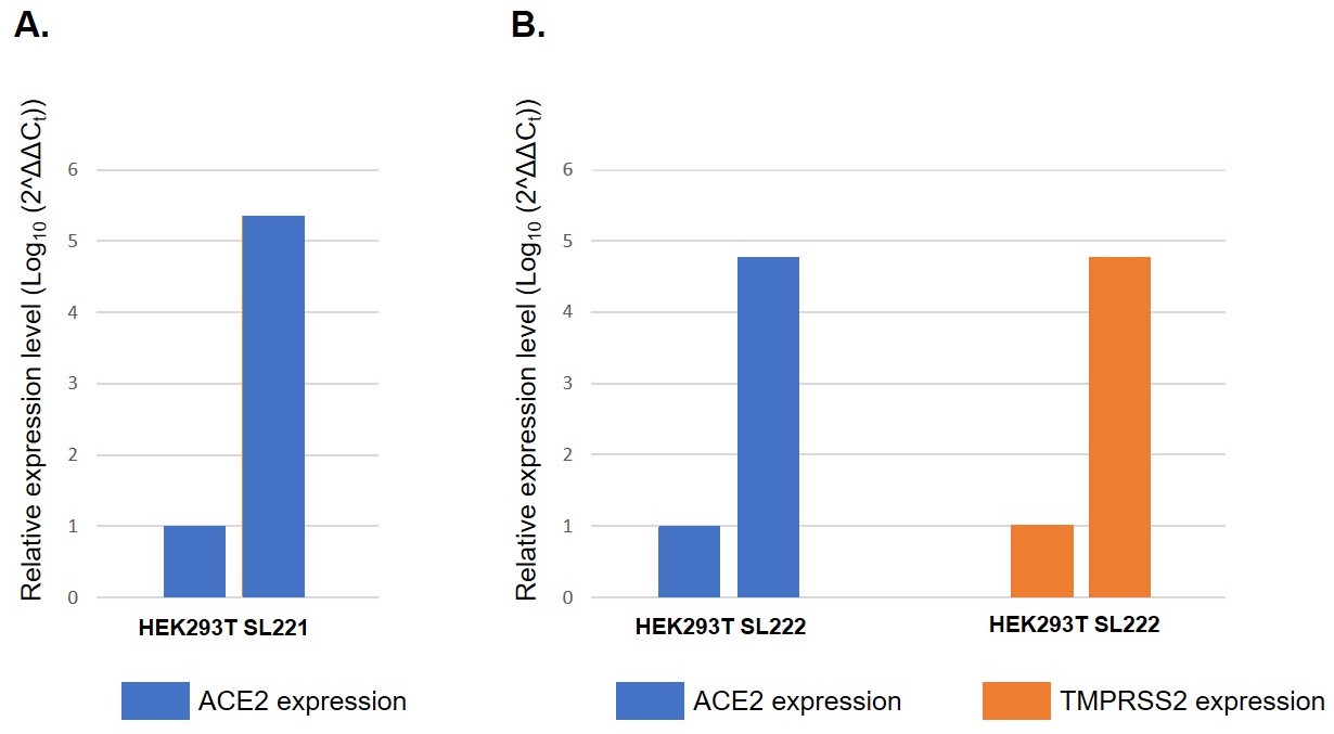
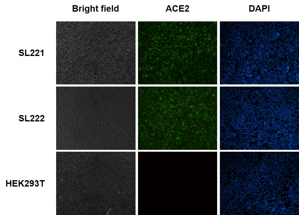
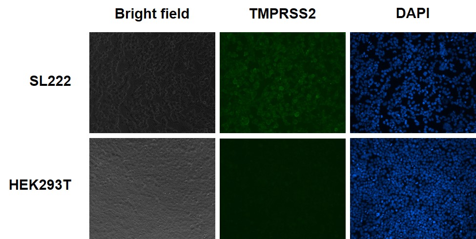
SL221 and SL222 are cell lines derived from HEK293T that express ACE2 alone or ACE2 and TMPRSS2 mRNA at high levels, as shown in Figure 1. Expression of ACE2 protein (Figure 2) and/or TMPRSS2 protein (Figure 3) on the cell surface makes these cells amenable to infection by Spike-pseudotyped lentivirus (Figure 4).
Figure 1. Relative mRNA expression levels of ACE2 and TMPRSS2 in GeneCopoeia stable cell lines. mRNA was amplified via RT-qPCR using primers specific for each gene. Ct values of ACE2 and TMPRSS2 were normalized to Ct values for housekeeping gene GAPDH. Relative expression levels were calculated from 2^ΔΔCt. A. ACE2 mRNA expression in parental cell line HEK293T and ACE2-overexpressing HEK293T cell line SL221. B. ACE2 and TMPRSS2 mRNA expression in parental cell line HEK293T and ACE2- and TMPRSS2-overexpressing cell line SL222.
Figure 2. Immunofluorescence (IF) analysis of ACE2 protein expression on the cell surface of ACE2-expressing HEK293T cells (Cat.# SL221), ACE2/TMPRSS2 expressing HEK293T cells (Cat.# SL222), and HEK293T cells. Unpermeablized cells were fixed, blocked and incubated with the primary anti-ACE2 antibody, then washed and incubated with fluorescent-labeled secondary antibody, washed again, and stained with DAPI.
Figure 3. Immunofluorescence (IF) analysis of TMPRSS2 protein expression on the cell surface of TMPRSS2-expressing HEK293T cells (Cat.# SL222) and HEK293T cells. Unpermeabilized cells were fixed, blocked and incubated with the primary anti-TMPRSS2 antibody, then washed and incubated with fluorescent-labeled secondary antibody, washed again, and stained with DAPI.
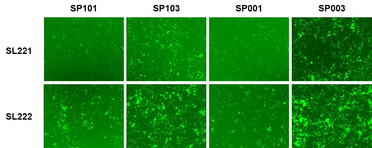
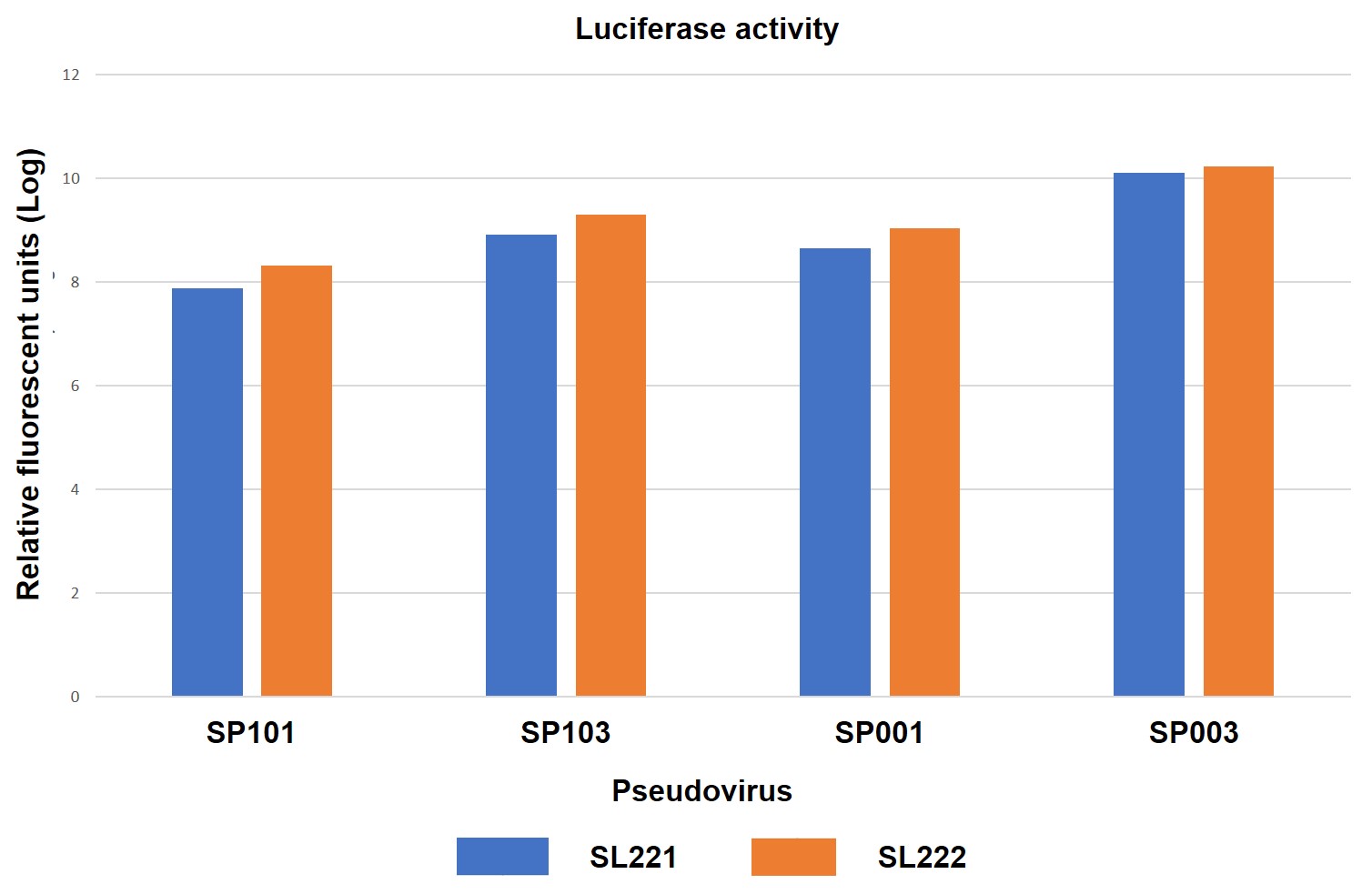
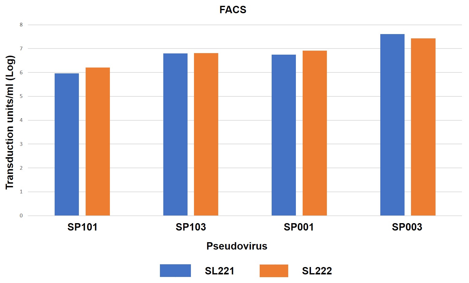
GeneCopoeia’s R&D team determined the ability of SARS-CoV-2 Spike-Pseudotyped Lentiviruses to infect ACE2-expressing cells, as shown in Figure 4.
A.
B.
C.
Figure 4.ACE2- or ACE2/TMPRSS2-expressing HEK293T cells transduced by eGFP/hLuc-expressing SARS-CoV-2 Spike pseudotyped lentiviruses (pseudoviruses). 10 μl of pseudovirus was used to transduce each cell line in a 96-well plate. 72 hours after transduction, cells were photographed under fluorescence microscopy, followed by measurement of luciferase activities or FACS sorting. SL221: HEK293T cells expressing ACE2. SL222: HEK293T cells expressing ACE2 and TMPRSS2. SP101: Full-length Spike protein, D614 variant. SP103: Full-length Spike protein, D614G variant. SP001: Truncated Spike protein, D614 variant. SP003: Truncated Spike protein, D614G variant. A. Fluorescence microscopy. B. Luciferase activity assay. 20 μl of cell lysate from pseudovirus transduced cell cultures were analyzed for firefly luciferase activity using GeneCopoeia’s Luc-Pair™ Firefly Luciferase HS Assay Kit (cat. no. LF007). C. FACS sorting for determination of titer in transduction units/ml.
Title: Advanced Tools For COVID-19 Coronavirus Research From GeneCopoeia
Presented Wednesday, September 9, 2020
The COVID-19 global pandemic, caused by the SARS-CoV-2 coronavirus, highlights an urgent need for diagnostic reagents, antiviral therapeutics, and vaccine development. In this webinar, we will discuss GeneCopoeia’s advanced solutions including testing kits, SARS-CoV-2 pseudotyped virus, and other tools needed for research into combating this deadly disease.
GeneCopoeia carries complete suites of state-of-the-art products for gene delivery using lentivirus and adenoassociated virus (AAV).
GeneCopoeia’s lentiviral solutions include:
Lentiviral plasmids. Available for expressing ORF cDNAs, gene promoters, miRNA precursors and inhibitors, CRISPR sgRNAs, and shRNAs for do-it-yourself, 3rd generation lentiviral packaging. Use the “Plasmids” tab for ordering information.
Ready-to-use particles. For infection of most mammalian cell types with either standard VSV-G pseudotyping, or for infection of ACE2-expressing cells with SARS-CoV-2 Spike glycoprotein pseudotyping. These particles can be produced in either in vitro or in vivo grade. See the lentivirus drop down menus below for ordering information.
Packaging kits. Enable you to package lentiviral transfer plasmids with either standard VSV-G pseudotyping or SARS-CoV-2 Spike glycoprotein pseudotyping. Use the packaging kits drop down menu below for ordering information.
GeneCopoeia’s AAV solutions include:
Ready-to-use particles. For infection of many mammalian cell types in nearly all available serotypes. These particles are be produced in in vivo grade, and can be used either in vitro or in vivo. Use the AAV drop down menu below for ordering information.
Serotype testing kits. For determining which AAV serotype(s) are best for infecting your cells of interest.
Lentiviral particles for SARS-CoV-2 viral protein expression are available for 2 primary applications:
Pseudotyping of lentivirus with the SARS-CoV-2 Spike (S) protein. Lentiviruses in common usage today are produced with the Vesicular Stomatis Virus G glycoprotein (VSV-G) expressed in the viral envelope, which enables the lentivirus to enter a wide range of mammalian cell types. However, during lentivirus packaging, the packaging plasmid encoding VSV-G can be replaced with a plasmid expressing SARS-CoV-2 Spike glycoprotein, which more narrowly limits their cell type infectivity to cells expressing ACE2, the human receptor for SARS-Cov-2 infection (Ou, X., et al., 2020. Nature Communications doi: 10.1038/s41467-020-15562-9; Hoffman, M., et al., 2020. Cell doi: 10.1016/j.cell.2020.02.052). These particles can be used for several applications, including vaccine development and testing antibodies that block cellular entry by SARS-CoV-2. If you want to order lentiviral particles pseudotyped with SARS-CoV-2 Spike protein, please contact inquiry@genecopoeia.com.
Expression of SARS-CoV-2 viral proteins from standard VSV-G pseudotyped lentivirus in stable cell lines.To order high titer, ready-to-use lentiviral particles expressing SARS-CoV-2 viral proteins in one of our more than 80 lentiviral vector types, select the product ID from the table below.
ORF10 protein [Severe acute respiratory syndrome coronavirus 2], full-length gene
*Note: Mature peptides are produced as a result of proteolytic processing of larger precursor proteins. To enable expression of these proteins in cells, an ATG has been added to the 5′ end of the ORF sequence.
Order AAV particles expressing SARS-CoV-2 viral proteins
To order AAV particles expressing SARS-CoV-2 viral proteins in one of out more than 250 non-viral and lentiviral vector types, select the product ID from the table below and contact inquiry@genecopoeia.com.
ORF10 protein [Severe acute respiratory syndrome coronavirus 2], full-length gene
*Note: Mature peptides are produced as a result of proteolytic processing of larger precursor proteins. To enable expression of these proteins in cells, an ATG has been added to the 5′ end of the ORF sequence.
Title: Advanced Tools For COVID-19 Coronavirus Research From GeneCopoeia
Presented Wednesday, September 9, 2020
The COVID-19 global pandemic, caused by the SARS-CoV-2 coronavirus, highlights an urgent need for diagnostic reagents, antiviral therapeutics, and vaccine development. In this webinar, we will discuss GeneCopoeia’s advanced solutions including testing kits, SARS-CoV-2 pseudotyped virus, and other tools needed for research into combating this deadly disease.

 Powered by Bioz