Select Page

qPCR arrays and reagents

Answer: With these arrays, you don’t need to spend time on primer design and validation of genes and miRNAs. These arrays allow you to perform batch qPCR detection of genes or miRNAs simultaneously in the same 96-well or 384-well plate. This greatly simplifies and speeds up your experiments.
Answer: GeneCopoeia offers custom services for miRNA and gene qPCR arrays that you have designed. You can visit our website and fill out the required information in your order. Please contact us directly if you have any questions.
Custom miRNA qPCR array order form
Custom gene qPCR array order form
Please contact us by email: inquiry@genecopoeia.com or call: 301-762-0888 and 301-762-0888.
Answer: Housekeeping genes are usually used as reference genes, but their expression is not fixed. The expression change of a particular housekeeping gene depends on the experimental conditions. The best method is to choose a series of common housekeeping genes, analyze their expression levels under different conditions, and then select a gene that is stably expressed.
Answer: Your choice of which RT primers to use depends heavily on the experimental design. If you use random primers, reverse transcription will begin at many different sites in the RNA. If you use oligo(dT), reverse transcription will start at the poly-A tail. In quantitative RT-PCR detection of specific genes, we recommend using both random primers and oligo(dT) in one reaction, in order to obtain higher efficiency of reverse transcription. In addition, you can use your own sequence-specific primer to prime reverse transcription in a one-step RT-PCR reaction.
Answer: The DNA polymerase used in these products is a special chemically-modified hot-start enzyme. Incubating for 10 minutes at 95℃ activates the enzyme.
Answer: This kit detects the expression of mature miRNA only.
Answer: The amplification product in miRNA qPCR is about 75bp. The melting temperature of the product is generally between 75℃ and 83℃.
Answer: Most of the All-in-One™ miRNA primers are designed to recognize the 3’-end of a template sequence. The matrue miRNA and the pre-miRNA have different sequences at the 3’-end of the base sequence. Therefore, the All-in-One™ miRNA primers will not amplify the pre-miRNAs.

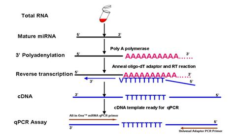
Answer: The experimental procedure includes two major steps (Figure 1). In the first step, polyA polymerase is used to add a polyA tail to the 3’ end of the miRNA. Simultaneously, MMLV reverse transcriptase, using a unique oligo(dT) adaptor primer, reverse transcribes the miRNA from the polyA tail. The second step, using the miRNA-specific forward primer and the Universal Adaptor primer as a PCR primer pair, along with a qPCR master mix containing SYBR® Green, specifically detects the reverse transcribed miRNA.


Figure 1. Experimental design used by the GeneCopoeia All-in-One™ miRNA qRT-PCR Detection Kit

Answer: A cDNA pool, containing reverse transcribed products from 10 human tissue total RNA samples, is used as the validation template. qPCR was performed using 0.2 uM primer and the GeneCopoeia All-in-One™ qPCR Mix. GeneCopoeia’s All-in-One™ qPCR pimers (or All-in-One™ miRNA qPCR primers) are validated to generate a single amplicon of the correct size for the targeted genes and miRNAs and to yield a single dissociation curve peak.
Answer: In combination with the All-in-One™ miRNA primers, our All-in-One™ miRNA qRT-PCR Detection Kit can detect miRNAs from as little as 10 pg of small RNA or 20 pg of total RNA.
Answer: PolyA polymerase adds poly-A tails to the 3’ end of all miRNAs, which can be reverse transcribed to first strand cDNA in the same tube. The resulting product can be used to detect the expression of any desired miRNAs by qPCR. By contrast, stem-loops are designed to detect just one or a few specific miRNAs, the 3’ end bases of which are paired with their sticky ends. Therefore, to detect such miRNAs, you must select a matching stem-loop. More than one RT reaction may need to be performed at the same time.
Answer: Total RNA has more types of RNA than small RNA, and so provides more templates to amplify in a qRT-PCR detection assay. This can help to improve the amplification sensitivity but could also reduce the amplification specificity.
Answer: The “RT” well contains a spike-in reverse transcription control, which can be used to monitor the efficiency of the reverse transcription reactions. The primer pair pre-loaded in the RT wells specifically amplifies a control cDNA template reverse transcribed from the spike-in exogenous RNA in the sample. The “PCR” well is a positive PCR control, which is used to verify the PCR efficiency by amplifying a pre-loaded DNA template with a specific pre-loaded primer pair. If the RNA sample is of high quality, the cycling program has been correctly run, and the thresholds have been correctly defined, the Ct value of the “RT” well should be 20±3, and the Ct value of the “PCR” well should be 20±2 across all arrays or samples.
Answer: The “GDC” well in each ExProfile™ gene qPCR array is a genomic DNA control. This is used to detect the presence of any genomic DNA contamination in each sample during each qPCR reaction. A Ct value for the genomic DNA control of lower than 35 indicates the presence of a detectable amount of genomic DNA contamination. Be sure to remove as much genomic DNA and residual contamination from your RNA samples as possible.

Stable Cell Line Services

Answer: There are two types of related services we currently offer in various cell lines. One consists of the generation of mammalian stable cell lines for protein expression, gene knockdown, CRISPR- or TALEN-mediated genome modifications, and primary cell immortalization. The second type is cell-based validation, in which we offer validation of ORF and cDNA expression levels, shRNA knockdown efficiency, CRISPR sgRNA validation, interaction between miRNAs and their miRNA targets, and promoter activity.

Answer: What is included in the cell-based service depends on the type of service you are interested in, as follows:

1. ORF expression. We will isolate either stable pools or single cell clones carrying stable integration of the ORF of your gene of interest. By default, we validate expression of the ORF by qPCR, although other validation methods (e.g. western, FACS, ELISA, etc.) are also available.

2. Knockdown by shRNA. We will isolate either stable pools or single cell clones carrying stable integration of the shRNA-expressing construct. By default, we validate downregulation of expression of the target gene by qPCR, although other validation methods (e.g. western, FACS, ELISA, etc.) are also available.

3. CRISPR genome editing. We offer virually any CRISPR-mediated applicaiton, including knockout, gene knock-in, mutagenesis, fusion tagging, gene activation, and gene repression. By default, we validate by sequencing the chromosome to detect the presence of the desired modification. Other validation methods (e.g. western, FACS, ELISA, etc.) are also available.

4. Primary cell immortalization. We typically offer immortalization of primary cells by infecting them with lentiviruses expressing either SV40 large T antigen, hTERT, or a combination of both. Using both SV40 large T antigen and hTERT together usually results in longer-lasting immortalization. We can also immortalize using other oncogenes, such as c-Myc, if desired. By default, we will check for expression of the immortalization gene by qPCR and to see if the cells are capable of fifteen or more generations (doublings). Other validation methods (e.g. western, FACS, ELISA, etc.) are also available.

5. Cell-based validation. We will provide a complete validation report containing the original test results and further analysis. The following methods are provided by default. Customers may also request other methods for validating or screening clones:


  • ORF expression validation: qRT-PCR, western blot and fluorescence microscopy (only for vectors with a fluorescence reporter gene)
  • shRNA: qRT-PCR
  • miRNA target validation: Luciferase reporter assay and fluorescence microscopy (only for vectors with a fluorescence reporter gene)
  • Promoter activity validation: Luciferase reporter assay or fluorescence microscopy (only for vectors with a fluorescence reporter gene)


Answer: For shRNA expression clones provided by GeneCopoeia, we guarantee that at least one in a set of three (3) shRNA constructs generates a knockdown efficiency of at least 70%. Customer-designed shRNAs do not carry this guarantee. Be aware that the knockdown efficiency of shRNAs depends on many factors, such as variability among different cell lines. We can identify the shRNA with the best knockdown performance in customer designated cell lines, and provide a detailed validation report. However, other shRNA constructs may work better in other cell lines. Therefore, if another cell line is needed in a future experiment, it is better for customers to also have shRNAs validated in that cell line for pre-screening.
Answer: We offer services in many cell lines such as HEK293, CHO, HeLa and NIH 3T3. However, if your desired cell line is not in this group, you may need to provide your cell line to us, or we might need to purchase it form a third party. After we perform quality control analysis, the project can be started. Please note that cell lines provided to us must be mycoplasma-free. We perform mycoplasma testing on all cells submitted to us or purchased from a third party..
Answer: This depends on what kind of vector you choose for your promoter clone. For example, our PG04 vector carries Gaussia Luciferance (Gluc) and Secreted Alkaline Phosphatase (SeAP), our PG02 vector carries Gluc, our PF02 vector carries GFP, and our PM02 vector is marked with mCherry. For the PG04 and PG02 vectors, we use the GeneCopoeia Secrete-Pair™ Dual Luminecence Assay Kit and the GeneCopoeia Secrete-Pair™ Gaussia Luciferace Assay Kit. We use fluorescence microscopy for the PF02 and PM02 vectors. We can validate your target promoter in a specific cell line for you as part of a complete service to support your research needs, sparing you much time and work.

Answer: Many plasmids have selectable markers like antibiotic resistance genes. We can introduce the genes of interest by plasmid transfection. Alternatively, if the desired cell line does not transfect well, we can use lentiviral transduction. Once the target gene is stably integrated into the host cell’s genome, antibiotics can be added to the growth medium to select for stable cell lines expressing the inserted gene. We can also use FACS to sort cells expressing fluorescent markers such as eGFP. Expression levels can be assayed by methods such as qPCR, western blot and ELISA. 


Genome editing products

General

Answer: If you are doing simple gene knockouts in humans, mice, or rats, you can order TALENs or CRISPR sgRNAs on our website. All you need to do is go to the TALEN/CRISPR search page, search for your gene, and then choose the appropriate clones that will work for your system. You can also order donor clones for these knockouts from the search results page. Please note that these TALENs and CRISPR sgRNAs are designed by default to target the protein coding-region as closely as possible to the initiator ATG of the splice variant (accession number) you select. This design strategy does not consider other possible variants of the gene. If the gene has multiple variants and you would like to target one particular exon, one unique variant, multiple variants, or all variants, or if you are doing a different application, such as introducing a point mutation, then you will need to contact us and, after determining what you need, we will send you a custom quote.
Answer: For sgRNA clones (including both all-in-one Cas9/sgRNA clones and sgRNA-only clones) as well as TALE clones, the default delivery format is bacterial stock. You have the option of ordering purified DNA for these clones for an additional charge. For HDR donor clones, the default delivery format is purified DNA. In addition, we offer ready-to-use lentiviral particles for Cas9 and sgRNA delivery.
Answer: The turnaround time for sgRNA clones (including both all-in-one Cas9/sgRNA clones and sgRNA-only clones) and TALE clones is 2-3 weeks. The turnaround time for HDR donor clones depends greatly on the nature of the modification that the clone is being used for. For HDR donor clones used for simple knockout, the turnaround time is 2-4 weeks. Other HDR donor clones, such as those used for fusion tagging or mutagenesis, can take 6-8 weeks, but can also take longer.
Answer: Yes. We sequence the inserts of each CRISPR sgRNA or TALE clone, and provide you with datasheets that show the full sequence of each clone (including HDR donor clones), a map, restriction enzyme digestions sites, and suggested sequencing primers. To obtain these datasheets, you just need to visit our datasheet download page on our website. You will need an account on our website, your catalog number(s), and your sales order number.
Answer: In the presence of drug, the only way for cells to survive is to integrate the plasmid into the chromosome, so it is possible to get drug-resistant clones that were transfected only with the donor plasmid. However, such integration is random. CRISPR increases donor targeting frequency by several orders of magnitude, and CRISPR-mediated donor integration is usually targeted.

Answer: Yes. For this you will need a donor that is homologous to your locus on either side of the base you want to mutate. The donor can be either a plasmid or a single-strand oligonucleotide. The donor is co-transfected with the genome editing tool (TALEN or CRISPR). Generation of a double strand break (DSB) leads to repair of the break by homology-directed repair (HDR) using the donor as a template.

GeneCopoeia recommends our donor plasmid design and construction service. We will construct a donor plasmid that contains a defined modification, flanked by a selectable marker such as puromycin resistance, and homologous arms from your target region. The donor may or may not also include a fluorescent reporter such as GFP. The markers can be flanked by loxP sites, to permit Cre-mediated removal, if desired. Use of a GeneCopoeia-designed donor plasmid allows you to select for edited clones and reduces the number of clones required for screening. You can also purchase our donor cloning vectors for do-it-yourself donor clone construction.

Answer: There are two strong reasons to use HDR for genome editing: 1) It is precise and controllable. Any desired change can be implemented; 2) You will be able to use a knocked-in marker (such as drug resistance or fluorescence), which will greatly facilitate your ability to identify candidate clones that have the modification you want. In addition, some applications, such as fusion tagging, require using HDR.

GeneCopoeia recommends our donor plasmid design and construction service. We will construct a donor plasmid that contains a defined modification, flanked by a selectable marker such as puromycin resistance, and homologous arms from your target region. The donor may or may not also include a fluorescent reporter such as GFP. The markers can be flanked by loxP sites, to permit Cre-mediated removal, if desired. Use of a GeneCopoeia-designed donor plasmid allows you to select for edited clones and reduces the number of clones required for screening. You can also purchase our donor cloning vectors for do-it-yourself donor clone construction.

Answer: Our genome editing products can be used for virtually all species. Our standard plasmids for both TALE  systems and CRISPR are designed for work in mammalian cells. In addition, these plasmids can be used as templates for T7 promoter-driven in vitro transcription, for introduction into mice, zebrafish, Drosophila, and many other model organisms. Further, we can generate custom constructs that can be used in a wide variety of organisms.
Answer: Yes. The donor must be present when the double strand break (DSB) is generated in order to be used as a repair template. Otherwise, the cell must use non-homologous end joining (NHEJ) to repair the DSB, because unrepaired DSBs are lethal.
Answer: Our TALE and CRISPR plasmids typically only integrate into the host genome at very low frequency in transfection experiments. However, after clonal selection for edited cells, we recommend screening clones for those which have lost the plasmids. This can be done by testing clones to see if they have become sensitive to the antibiotic of the resistance gene on the plasmid, or if they no longer express the plasmid’s fluorescent marker (where applicable). Our lentiviral clones, when packaged into lentiviral particles and used to infect cells, are expected to integrate randomly into chromosomes.

Answer: The presence of the CMV or other promoters driving expression of the Cas9 or TALE coding sequences permits expression from the plasmid DNA. sgRNA transcription is driven by the U6 promoter. We recommend that you use the most efficient method for your cell type of interest. The following are all acceptable approaches for delivering our genome editing tools into cells:

1. Standard transfection. GeneCopoeia recommends our EndoFectin™ transfection reagents. Transfection can be used for either DNA, RNA, or Cas9 nuclease protein.

2. Electroporation, which can be used for either DNA, RNA, or Cas9 nuclease protein.

3. Lentiviral transduction (cannot be used with HDR donors).

4. Micro-injection of either DNA, mRNA, .or Cas9 nuclease protein. The plasmids can be used as templates for in vitro transcription of RNA, or synthetic RNA can be used.

Answer: If you are using TALEN or CRISPR to create mutations in your gene of interest without the use of a homologous donor, then you will need to undertake much time-consuming, labor-intensive downstream work in order to identify edited clones. After transfection, you will need to isolate many colonies grown up from single cells. Next, the screening procedure depends on the nature of the modification, as described below:

1. If you are making an insertion or deletion, the easiest way to screen your cells is by PCR using primers flanking the modified site, provided that the insertion or deletion is large enough to detect by standard agarose gel electrophoresis.

2. For very small insertions or deletions, you can screen your clones using GeneCopoeia’s IndelCheck™ T7 endonuclease I assay, which is a method that detects mutations by cleaving double stranded DNA containing a mismatch. You can also screen using Sanger sequencing of PCR products. Ultimately, Sanger sequencing needs to be done to verify the presence of the modification.

3. If you are introducing a point mutation, then you can use either real-time PCR or Sanger sequencing to detect the modification.

4. If the modification you are introducing creates or destroys a restriction enzyme site, then enzyme cleavage of PCR products can be used to distinguish between modified and unmodified alleles.

5. Finally, either Sanger sequencing of PCR products or Next Generation sequencing of whole genomes can be used to screen for modifications. Regardless of which screening method you choose, it is also important that you are able to determine whether only a portion or all of the alleles have been modified.

In order to reduce the amount of time and effort required to identify edited clones, GeneCopoeia recommends our donor plasmid design and construction service. We will construct a donor plasmid that contains a defined modification, flanked by a selectable marker such as puromycin resistance, and homologous arms from your target region. The donor may or may not also include a fluorescent reporter such as GFP. The markers can be flanked by loxP sites, to permit Cre-mediated removal, if desired. Use of a GeneCopoeia-designed donor plasmid allows you to select for edited clones and reduces the number of clones required for screening. You can also purchase our donor cloning vectors for do-it-yourself donor clone construction.

Answer: For safe harbor integration, we recommend PCR using primers designed to amplify the recombination loci, or Southern blotting, which can rule out random integration. All GeneCopoeia Safe Harbor Gene knock-in Kits contain PCR primer pairs intended for this exact purpose. For other editing experiments using HDR, junction PCR can be be used to identify edited sites. The junction PCR method will will only yield product if donor integration occurred at the correct genomic site. Southern blotting or PCR using primers recognizing the plasmid backbone can also be used to rule out random integration.
Answer: Yes. Even though frameshifts are not possible with miRNAs and other noncoding RNAs, an indel occurring in a critical region, such as the mature sequence of a miRNA, should be enough to abolish its function.
Answer: The vector backbones of our TAL effectors or CRISPR sgRNAs are designed to not replicate in the host. These plasmids, which are transiently transfected, will typically be lost after several rounds of cell division and will not further affect the host cell. After transfection, cells are plated at low density to promote the formation of single colonies. These colonies should be screened to ensure that they have lost the plasmid(s). This can be done by testing clones to see if they have become sensitive to the antibiotic of the resistance gene on the plasmid, or if they no longer express the plasmid’s fluorescent marker (where applicable). However, even if the TALEN or CRISPR plasmid integrates, it can no longer cut the site after it is edited, because NHEJ destroys the TALEN or sgRNA recognition site. To be completely assured that the transfection is transient, we recommend delivering RNA or Cas9 nuclease protein instead of plasmid DNA. If you are using HDR, we recommend engineering synonymous mutations into the donor to destroy the TALEN or sgRNA recognition site without changing the amino acid sequence.
Answer: Yes. Both TALEN and CRISPR have been shown to be able to disrupt multiple copies at once. The efficiency varies depending on different factors, such as cell type, transfection efficiency and TALEN/CRISPR activity.

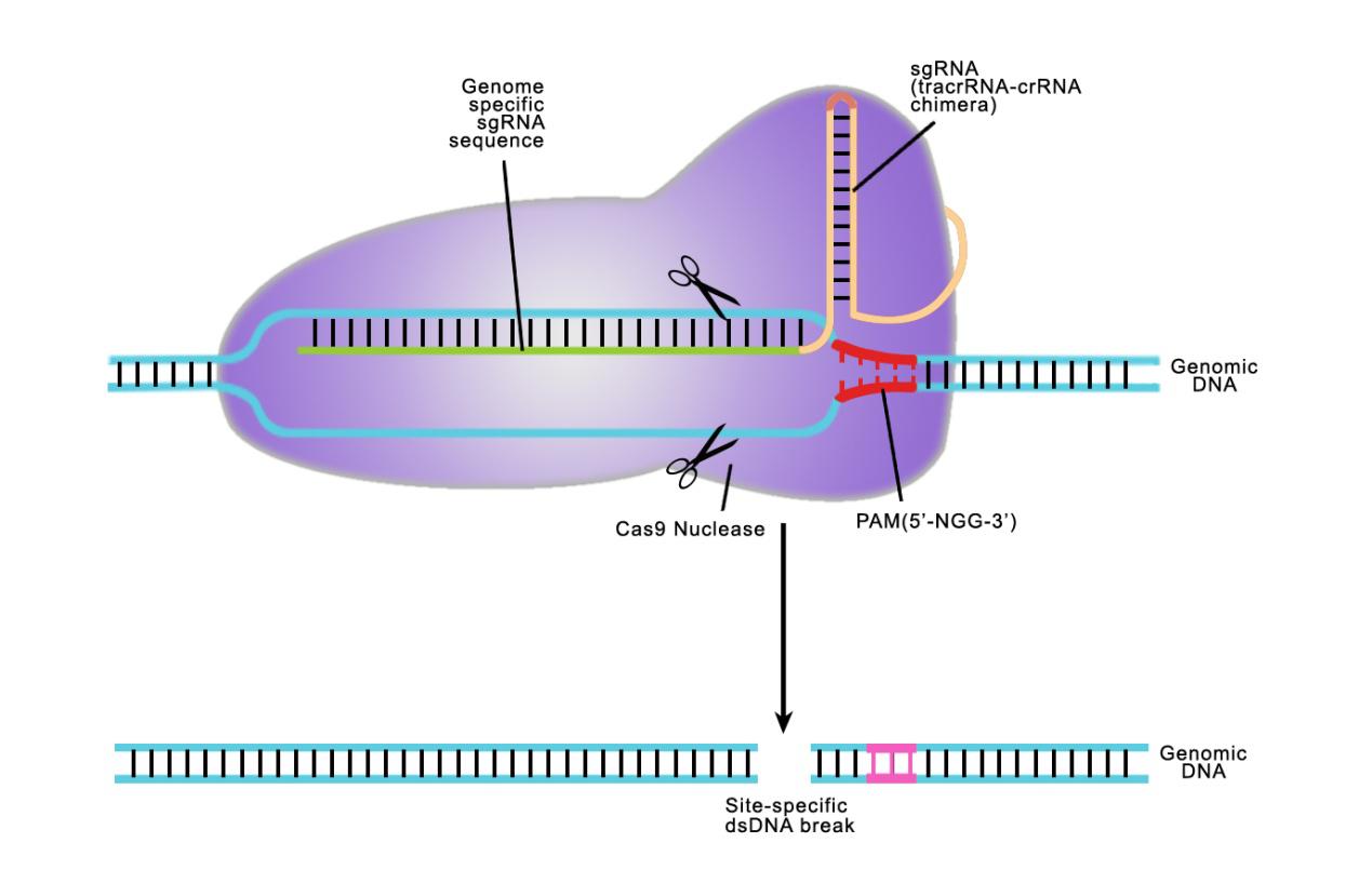
Answer: Both TALE and CRISPR recognize a specifi target sequence to initiate genome editing. The biggest difference is in how they do it. CRISPR uses a single guide RNA (sgRNA) homologous to a 20 nucleotide target sequence. This target sequence must be immediately followed by a 3 nucleotide N-G-G sequence known as the Protospacer Adjacent Motif (PAM). The single guide RNA guides the Cas9 nuclease to the desired site. When using the CRISPR system, we need to design the sequence of sgRNAs.
TALEs are proteins that recognize target sequences using variable amino acids in a series of 34 amino acid repeats. These variant amino acids are known as Repeat Variable Diresidues (RVDs). Each repeat contains one RVD, and each of the RVDs recognizes a specific nucleotide.
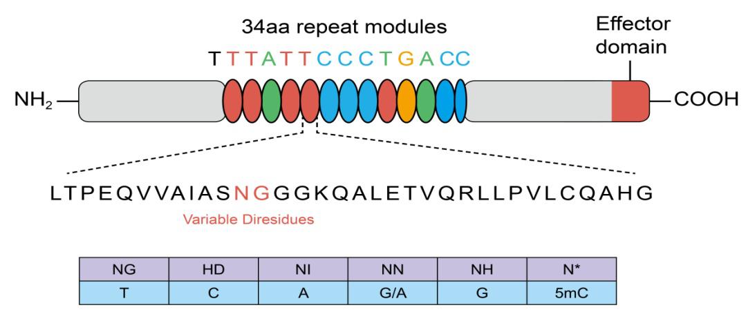
TALEs are fused to their genome editing motifs, which can include nucleases or transcriptional modulators. When using TALE technology systems, we need to assemble RVDs in the correct order to match the target sequence.

Answer: There is never an easy answer to this question. It all depends on what you are trying to accomplish. Each system has its advantages and disadvantages. CRISPR tends to edit genes at higher efficiency than TALEN. Also, CRISPR is not sensitive to DNA methylation, and is much more amenable to multiplexing, giving it further advantages over TALEN. On the other hand, CRISPR, including the Cas9 nickase, is more prone to modifying off-target sites, which, depending on your particular application may or may not be an issue. There are also “high-fidelity” variants of Cas9 nuclease that edit genes with greater specificity, but sometimes with reduced efficacy and with increased design constraints.


TALE

Answer: Yes.


CRISPR

Answer: Yes. We have the reagents for the Cas9 D10A nickase, and have successfully tested our double nickase designs. However, in order to create mutagenic DSBs, the nickase requires the correct targeting of two appropriately-spaced sgRNAs on opposite strands, flanking the break site. Because proper sgRNA targeting requires the presence of the N-G-G “PAM” site immediately following the recognition site, it might not always be possible to use the nickase for DSB formation. There are also “high-fidelity” variants of Cas9 nuclease that edit genes with greater specificity than wild type Cas9, but sometimes with reduced efficacy and with increased design constraints. However, since these high fidelity variants use only one sgRNA, they are easier to work with than Cas9 nickases. In addition, using Cas9 nuclease protein instead of a DNA plasmid could help reduce off-target activity.
Answer: Yes. To create a DSB, the nickase requires the correct targeting of two appropriately-spaced sgRNAs on opposite strands, flanking the break site. This is sufficient to stimulate HDR between the target site and the donor. While this method has the advantage of potentially fewer off-target NHEJ-mediated mutations, since single strand nicks are repaired with higher fidelity than DSBs, it is not without limitations. Proper sgRNA targeting requires the presence of the N-G-G “PAM” site immediately following the recognition site. Therefore, it might not always be possible to use the nickase for HDR. There are also “high-fidelity” variants of Cas9 nuclease that edit genes with greater specificity than wild type Cas9, but sometimes with reduced efficacy and with increased design constraints. However, since these high fidelity variants use only one sgRNA, they are easier to work with than Cas9 nickases. In addition, using Cas9 nuclease protein instead of a DNA plasmid could help reduce off-target activity.
Answer: Yes. We offer Cas9 nuclease protein that can be used to form a ribonucleoprotein (RNP) complex with either synthetic or in vitro transcribed sgRNA. The RNP can then be used to transfect cells with our CRISPR-Fectin™ transfection reagent.
Answer: We only sell plasmids containing our custom-designed CRISPR sgRNAs. If you need a negative control, we also sell CRISPR plasmids containing a scrambled sgRNA.
Answer: Yes.
Answer: Yes. There is a double mutant of the Cas9 nuclease that completely abolishes nuclease activity. This mutant can be fused to a transcriptional modulator such as VP64 and targeted to specific genes. You can also use the catalytically dead Cas9 with properly-designed sgRNAs to repress, or interfere with, gene expression. For more information, please visit our pages on CRISPR activation (CRISPRa) and CRISPR interference (CRISPRi).
Answer: Yes. We have both non-viral and lentiviral formats. We also have custom lentiviral particle production services, in which we can provide you with lentiviral particles expressing both Cas9 and sgRNAs.
Answer: Unfortunately, no. Lentiviruses enter cells as RNA, but HDR donors must enter the cells as DNA at the same time as Cas9 and the sgRNAs.
Answer: Cas9 stable cell lines are primarily useful for lentiviral CRISPR delivery and CRISPR sgRNA library screening. sgRNA library screening is typically done with lentivral delivery. In principle, it is possible to either co-infect your cell line with one Cas9-expressing lentivirus and with an sgRNA-expressing lentivirus, or with a single “all-in-one” lentivirus (Cas9 and sgRNA expressed in the same virus). However, in practice, due to the large size of the Cas9 gene (4.4 kb), the titers of Cas9-expressing lentiviruses tend to be much lower than sgRNA-expressing lentiviruses. Therefore, we recommend using a cell line that has previously had Cas9 stably integrated in the genome for infection with lentiviral-based sgRNA libraries. GeneCopoeia carries a large collection of pre-made Cas9-expressing stable cell lines, as well as stable cell lines expressing the machinery for CRISPR activation (CRISPRa) and CRISPR interference (CRISPRi).  You can also purchase reagents that you can use to build a Cas9 stable cell line yourself, or you can have GeneCopoeia create a custom Cas9-expressing stable cell line as a custom cell service project.


sgRNA libraries

Answer: Lentiviral particles, transfection-ready DNA, and bacterial stock.
Answer: Yes. Our sgRNA libraries are available standard as pools. However, if you wish to receive your libraries as individually arrayed clones, simply contact us for a custom quote.
Answer: Yes. However, we strongly recommend that you use a cell line that is stably expressing Cas9 to transduce sgRNA-expressing lentiviral particles with, in order to best maintain consistent representation of each sgRNA in the library. To save you time and effort, GeneCopoeia provides a large collection of Cas9-expressing stable cell lines that are ideally suited for sgRNA library screening. If you would like to create a Cas9-expressing stable cell line yourself, you should first transduce your cells with Cas9-expressing lentiviral particles. Once you have established the stable Cas9-expressing cell line, use the sgRNA libraries for transduction.
Answer: Yes. The lentiviral plasmids are “dual-use”, so that they can either be packaged into lentiviral particles or transfected into cells by standard transfection methods.
Answer: Our sgRNA representation does not need to be validated by Next Generation Sequencing. Each library is small compared with the genome-wide libraries, and each sgRNA clone is constructed individually, cultured in E. coli individually, then pooled as E. coli in approximately equal amounts. From those pools we prepare DNA and then, if necessary, lentiviral particles.

shRNA clone sets

Answer: All you need to do is go to the shRNA clones search page, search for your gene, and then choose the appropriate clones that will work for your system. If you have any custom requirements, then you will need to contact us and, after determining what you need, we will send you a custom quote.
Answer: GeneCopoeia provides a set containing 3 individual shRNA constructs and a scrambled control. Each clone is provided as of 5 µg purified plasmid. A datasheet containing information for each clone, vector and restriction enzyme sites is available for download on our website. You will need a GeneCopoeia account, your sales order number, and your catalog number(s) in order to download the datasheet.
Answer: GeneCopoeia guarantees that the shRNA sequences in the expression cassettes are identical to the target gene. If none of the four constructs produce a 70% or greater knockdown efficiency as determined by qRT-PCR, and the inefficiency is caused by a flaw in our construct design, then we will provide another set of four new clones targeting the specific gene free of charge.
Answer: We offer both promoters so customers can choose based on their preference. U6 is the stronger promoter, however, because it is stronger there is a greater chance of causing off-targeting effects or cellular toxicity. In most cases either promoter should be fine to use, however, there are tissue and cell specificities associated with each promoter. We recommend doing a literature search to see what other researchers have used for the particular cells you are working with.


Answer: Yes, Stbl3 is recommended.
Answer: You should measure the knockdown efficiency when the transfection efficiency is greater than 80%. The reporter gene in the vector is used to monitor transfection efficiency. Alternatively, if the transfection efficiency of your cell line is low, you can order the lentiviral versions of your clones and transduce your cells with the lentiviruses. RNA can be harvested from transfected cells and used in quantitative RT-PCR to estimate the reduction in gene expression. Western blot is recommended over qPCR to evaluate the silencing effect of the shRNA construct(s). Gene expression levels from cells transfected with a scrambled control clone must be compared with the shRNA transfected samples.
Answer: Factors influencing transfection efficiency include 1) The quality of the plasmid DNA; 2) The condition of the cells; 3) The quality, condition, or age of your transfection reagents and plasmids; 4) The cell density at the time of transfection; and 5) The contact time between cells and the DNA/transfection reagent complex.
Answer: The scrambled control clone is constructed by cloning a scrambled sequence (one that does not match any genomic sequences) into shRNA vectors. It serves as a negative control to eliminate the potential non-specific effect induced by expression of the plasmid.
Answer: We strongly recommend performing a kill curve on each batch of cells to determine the optimal puromycin concentration.

Lentivirus

Answer: The best way is to search for what you need on our lentiviral search page. You simply need to indicate the clone type (ORF, promoter, etc.), the gene information (gene symbol, aliases, description, nucleotide accession, Entrez gene ID, catalog or product ID), the vector type, and the delivery volume. You can also request a custom quote by contacting us.
Answer: 50 μl, 100 μl, 200 μl, and 400 μl, at titers of 10^7-10^9 TU/mL.
Answer: You can use lentivirus to infect or transduce virtually any mammalian cell type. The original HIV genome, from which lentiirus is derived, has been modified over several generations to make it a safer and more useful gene delivery vehicle. In the 3rd generation lentiviral vectors that GeneCopoeia uses, the HIV envelope (env) glycoprotein, which is necessary for infection of CD4+ T-cells, has been replaced with the vesicular stomatitis virus G (VSV-G) glycoprotein. VSV-G enables lentiviruses to infect virtually all mammalian cell types.
Answer: Lentiviruses comprise a subtype of retroviruses. Lentiviruses can stably integrate into the host genome in dividing, non-dividing and post-mitotic mammalian cells, while retroviruses are less active in this scenario. Adenoviruses can also transduce non-dividing cells, but can’t stably integrate into the host cell’s genome. Adenoviruses also take much more time to design and prepare. In addition, lentiviruses are much less immunogenic than the retroviruses and adenoviruses, making lentivirus more suitable for use in most types of cells and animal models.
Answer: One of the key factors of a successful transduction is the cell type. For example, transduction efficiency is much higher in actively dividing cells than in non-dividing cells. In addition, transduction of cells works better at lower MOI (multiplicity of infection) than at higher MOI. MOI is the ratio of the number of lentivirus particles to the number of cells. For some cell types, the higher the MOI , the larger the volume and higher the titer of lentivirus is required in order for the experiment to succeed. You can adjust the cell number and add the appropriate amount of lentivirus according to what has been reported in the scientific literature. If there is no adequate information in the scientific literature, we recommend performing a preliminary experiment using gradient dilutions of lentivirus, such as 0.1 μl, 0.3 μl, 0.5 μl, 0.7 μl, 0.9 μl for GeneCopoeia purified particles. Another important consideration for getting good transduction efficiency is the cell status. Transduction efficiency varies greatly between healthy cells and unhealthy cells. Therefore, it is essential to keep the cells as healthy as possible. For some cells with high MOI, you could also include additives such as polybrene to enhance the transduction efficiency. However, the overall health of the cells itself is always the most essential element.
Answer: Before lentivirus production starts, you need to first prepare the plasmid DNA using a well-established purification method. Make sure the plasmid you prepare is of the highest possible quality. You can measure its purity by the absorption ratio of 260 nm to 280 nm. You should also check the integrity of the plasmid by agarose gel electrophoresis. GeneCopoeia can provide your clones in transfection-ready format. In rare cases, toxic genes and large fragment inserts lead to low titers. Next, ensure your lentiviral packaging cell line is well maintained and passaged regularly, and make sure the culture is free from contamination of bacteria, fungi, and/or mycoplasma. Further, use an optimized lentivirus packaging system and reagent. We recommend the GeneCopoeia Lenti-Pac™ HIV-Based Lentiviral Packaging system, which is optimized for production of high viral titer, together with the GeneCopoeia Lenti-Pac™ HIV Expression Packaging Kit. We also recommend using GeneCopoeia 293Ta lentiviral packaging cells (Cat No. LT008). These cells are guaranteed to provide higher transfection efficiency and lower cell toxicity.

Answer: There are two main GOI-related factors that can affect the lentiviral titer. These are:

Toxicity. Some genes expressed from lentiviruses are toxic to cells and so will sharply reduce the titer.

Insert length. Larger inserts also tend to reduce viral titer.

The issue of insert length must be considered along with the backbone vector size, because the viral titer is influenced by the total length of the plasmid. Our recommendations for insert size for lentivirus packaging are shown below. Note that these are only general guidelines:

For Lv200 series vectors (such as Lv201 and Lv206), the insert should be less than 4 Kb.

For most Lv100 series vectors, except for those with eGFP fusions, the insert should be less than 5kb.

For other stripped-down vectors (without reporter or selection gene), the insert plus HIV related parts in the packing vector should be less than 10 Kb.

Answer: In general, a lentivirus packaging system contains a gene transfer vector plasmid and a packaging plasmid. The second and third generation lentivirus packaging systems are both designed to separate the essential genes of the transfer vector, envelope and packaging components onto different plasmids, thereby reducing the risk of recombination. When some lentiviral structural proteins must be expressed along with the gene of interest in the second generation, the vector of the third generation is revamped to make it self-inactivating and tat-independent. The GeneCopoeia™ HIV-Based Lentiviral Expression System is a modified version of the third generation self-inactivating (SIN) lentiviral vector system, which incorporates enhanced biosafety features and is optimized for production of high viral titers.
Answer: The titer of the GeneCopoeia lentivirus products in each lot are determined by qRT-PCR, which shows the physical number of viral genomic RNA molecules. The numerical relationship between titer (physical copy number) and transduction unit (TU or IFU) can be basically summarized using the following formula: TU= Titer (physical copy number)/100. The titer may vary among different cell types. In general, we recommended conducting a preliminary experiment before your formal study to ensure the viability of the lentivirus stock and to test the amount needed to transduce the cell type of interest. For more information regarding titer estimation by transduction, you may download a copy of the user manual.
Answer: It is safe to use GeneCopoeia lentivirus. The GeneCopoeia HIV-Based Lentiviral Expression System meets Biosafety Level 2 (BSL-2) requirements based on the criteria published by the Centers for Disease Control and Prevention (CDC). This system is a modified version of the third generation of the self-inactivating (SIN) lentiviral vector system, which incorporates enhanced biosafety features. The lentiviral transfer vector is responsible for transduction and stable integration into the genome of the host cell, but lacks the elements essential for transcription and packaging lentiviral particles by itself. Thus, it is self-inactivated, meaning that no unwanted viral replication and production will happen after the first transfection. Nevertheless, the guidelines for working with BSL-2 safety category materials must be adhered to. For more information regarding BSL-2, please visit the CDC website.
Answer: GeneCopoeia offers a vast number of clones in a HIV-based lentiviral system, which includes more than 40,000 human and mouse ORF expression clones, small hairpin RNAi (shRNA) against genome-wide target genes from human, mouse, rat and other animals, miRNA inhibitor clones for all known human, mouse and rat miRNAs, promoter reporter clones for more than 20,000 human and 18,000 mouse promoters, and CRISPR sgRNA clones for human and mouse. This system provides high expression levels and high efficiency of gene delivery to virtually all mammalian cell types. The lentiviral expression construct was validated by full-length sequencing, restriction enzyme digestion, and PCR-size validation using gene-specific and vector-specific primers. Together with GeneCopoeia’s EndoFectin Lenti Reagent (Cat No. EF001), TiterBoost™ reagent, 293Ta lentiviral packaging cells (Cat No. LT008) and MycoGuard™ mycoplasma detection kit, GeneCopoeia’s lentiviral products provide high viral titer and are confirmed free of bacteria, fungi and common mycoplasma contamination.
Answer: Lentivirus has some level of toxicity to cells. It may cause damage to your cell of interest with either superfluous amounts of lentivirus, or if the infection were allowed to go on for too long a period of time. In these cases, you can adjust the multiplicity of infection (MOI) to a lower range. We recommend replacing the old culture medium with fresh complete medium 4-8 hours post transduction (no later than 12 hours post transduction).
Answer: Lentiviruses can stably integrate into the host cell’s genome and obtain a consistent level of expression. With a selectable marker in the lentiviral gene transfer vector plasmid, it is easy to generate a stable cell line using drug selection. You can use qRT-PCR, western blot or other detection methods to estimate the expression level of your gene.