Introduction
Click chemistry describes a class of chemical reactions that use bio-orthogonal or biologically unique moieties to label and detect a molecule of interest in mild, aqueous conditions. DBCO alkynes can be used to perform click reactions with azide-modified targets without the use of heavy metal catalysis. DBCO reactions are ideal for surface labeling of live cells and also minimize damage to fluorescent proteins like GFP or R-PE.
The Cy5 DBCO is reactive with azide via a Strain-promoted Azide-Alkyne Click Chemistry reaction (SPAAC).
Features
Efficiency—the click reaction is complete in less than 1 hour;
Specificity—the reaction between the label and detection tag is selective and specific;
Stability—the reaction product contains an irreversible, covalent bond;
Biologically inert—the components of the reaction do not undergo any side reactions.
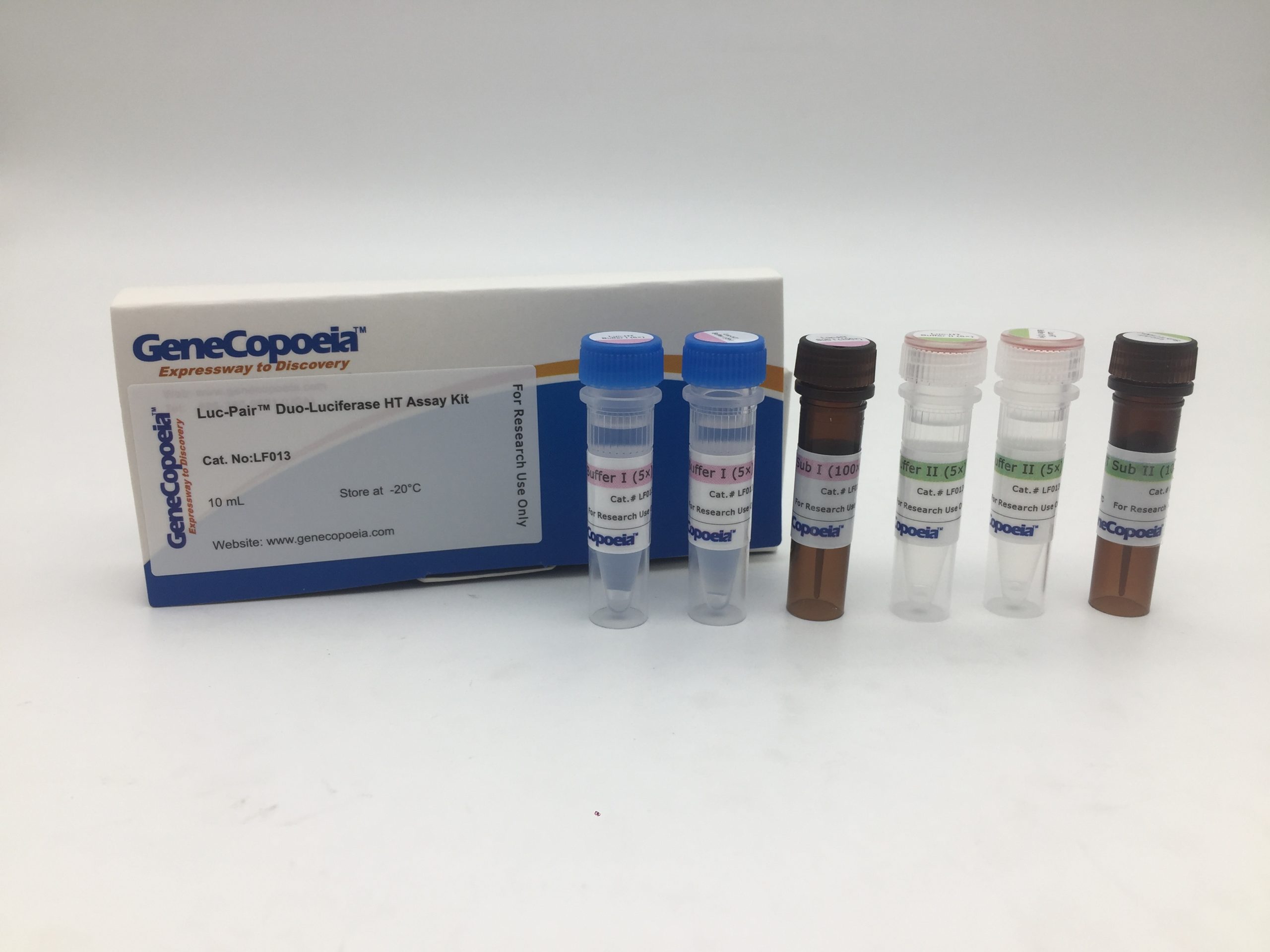
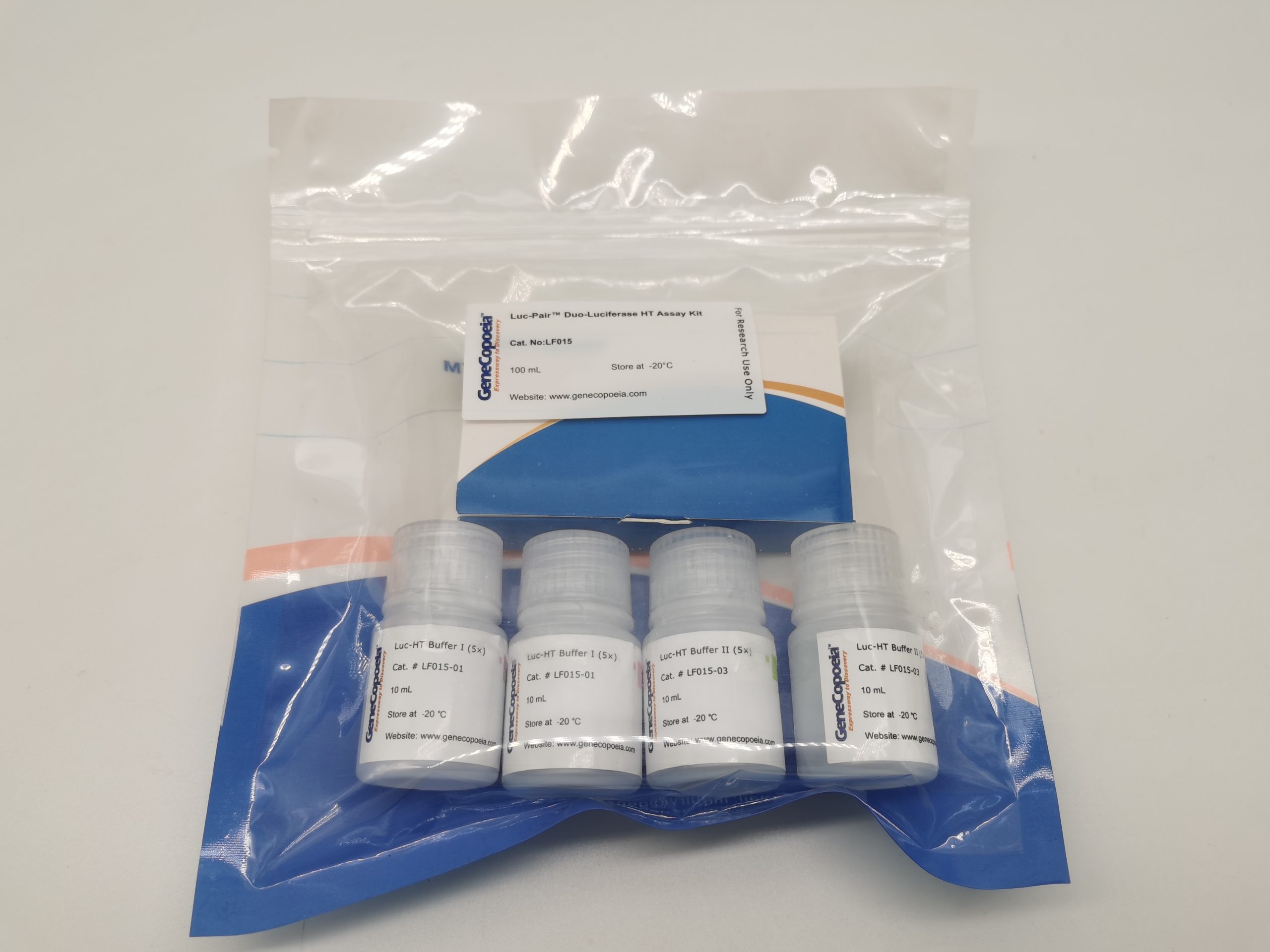
GeneCopoeia’s Luc-Pair™ Duo-Luciferase HT Assay Kit is a convenient system for high-throughput detection of Firefly luciferase (FLuc) and Renilla luciferase (Rluc) activities in 96- or 384-well plates. The Luc-Pair™ Duo-Luciferase HT Assay reagents can be added directly to cells in growth medium without washing or preconditioning, and are optimized for use with many different media containing 0–10% serum (e.g. DMEM, RPMI1640, etc.) Firefly luciferase luminescence is produced by one reagent, while a second reagent simultaneously quenches the Firefly luciferase and produces Renilla luciferase luminescence.
The Luc-Pair™ Duo-Luciferase HT Assay Kit works great for many different eukaryotic cell types (e.g. mammalian, invertebrates, etc.) and is compatible with GeneCopoeia, Promega and many other Fluc and Rluc vectors.
Advantages
· Enhanced stability. · Convenient — Directly lyse cells in culture medium and measure luciferase activities simultaneously. · Versatility — Works well with many different eukaryotic (adherent or suspended) cells using micro-plate luminescence readers. · Low background — Very limited background luminescence. No subtraction from readings is required. · Simplicity — Renilla luciferase buffer contains the quenchers for Firefly luciferase activity (Figure 3). enabling a quick Glow and Stop-N-Glow two-step assays. · Reproducibility — Reliable, linear results for a concentration range over several orders of magnitude.
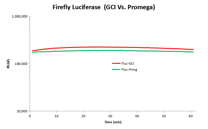
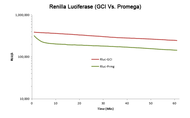
Figure 1.Activities of Firefly and Renilla luciferase signals using the GeneCopoeia (GCI) Luc-Pair Duo-Luciferase HT Assay Kit. HEK 293 cells were transfected with Promega pGL4.13/pGL4.75 reporter vectors for 48 hours. FLuc and RLuc activities were measured as described in the procedure. Promega’s Dual-Glo luciferase assay kit was used (FLuc-Prmg and RLuc-Prmg) in comparison.
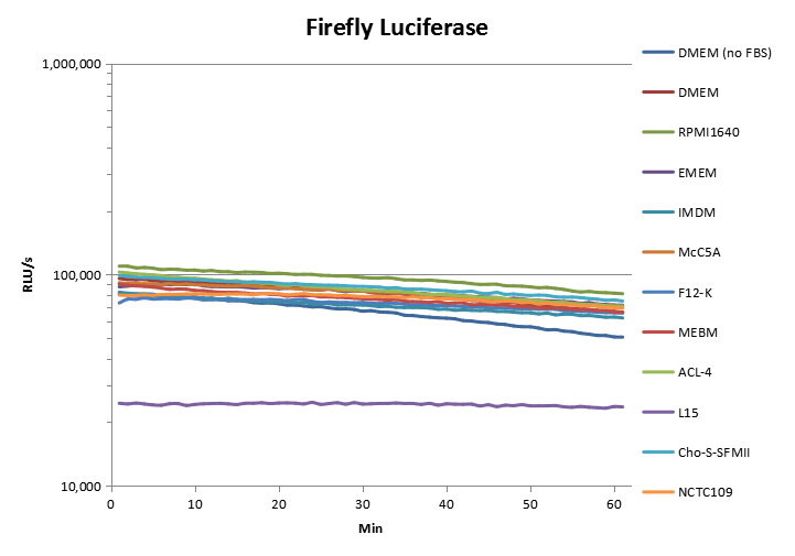
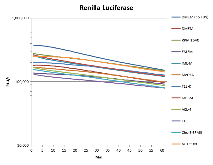
Figure 2.Firefly and Renilla luciferase activity mesured in various media. HEK 293 cells were transfected with Promega pGL4.13/pGL4.75 reporter vectors for 48 hours. 5×10⁴ transfected cells were suspended in DMEM without serum or the following types of media containing 10% serum: DMEM, RPMI1640, EMEM, IMDM, McCoy’s 5A, F-12K, MEBM, ACL-4, L15, Cho-S-SFMII, NCTC109. Firefly and Renilla luciferase activities were measured as described in the procedure.
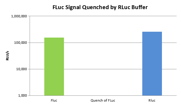
Figure 3. Firefly luciferase activity is quenched with the addition of Luc-HT Buffer II. HEK 293 cells were transfected with Promega pGL4.13/pGL4.75 reporter vectors for 48 hours. Firefly luciferase activity was measured as described in the procedure. Next, 1× Luc-HT Buffer II (without or with substrate) was added to the wells, followed by count reading in a luminometer. About 99.9% of firefly luciferase activity was quenched (middle column).
*Promotions are valid in the US & Canada only. For international customers, please contact your local distributors. Discounts are not valid on previous purchases and cannot be combined with additional discounts.
Answer: To prevent loss of activity, it is best to avoid repeated freezing and thawing. We recommend pipetting small aliquots into new, clean, sterile tubes and placing those aliquots at -20°C. Once you have thawed one of the aliquots, discard it instead of putting it back in the freezer.
Answer: The Gaussia luciferase substrate, coelenterazine, is also the substrate for Renilla luciferase, so it is compatible with Renilla. However, coelenterazine cannot be used with Firefly (Photinus pyralis) luciferase, which instead uses luciferin as its substrate.
GeneCopoeia’s AAVPrime™ Adeno-associated virus (AAV) products are the ideal tools for expressing or modifying genes in a broad range of cell and tissue types, especially in vivo, with high efficiency and enhanced safety. GeneCopoeia’s optimized helper-free human AAV system allows viral packaging without potentially pathogenic helper adenovirus. Many pre-made AAV are available in 25 different serotypes, such as fluorescent reporters. You can also request us to produce custom AAV for genes up to 3 kb in length.
High titers. Titer of purified AAV particles can be up to 1014 GC/ml.
High adaptability. Packaging types include-ORF cDNA, CRISPR, shRNA, or miRNA.
Low toxicity. Does not integrate into the host genome.
Low immunogenicity. Minimal host immune response.
Safe. Not associated with any human disease
Search all AAV products by gene ID or accession number
*Promotions are valid in the US & Canada only. For international customers, please contact your local distributors. Discounts are not valid on previous purchases and cannot be combined with additional discounts.
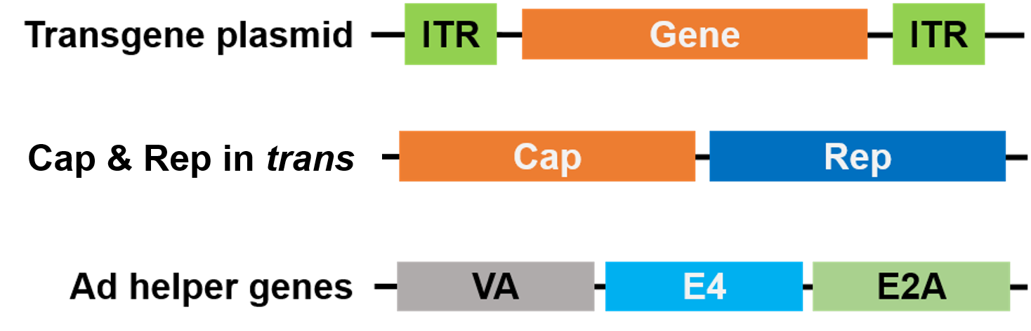
AAVPrime™ uses a helper virus-free system for the safe preparation of high titer AAV. HEK293T cells are co-transfected with three plasmids encoding the factors necessary for recombinant AAV packaging. The AAV particles are then either membrane purified or purified by two-phase partitioning for high-titer AAV ready for in vivo animal use.
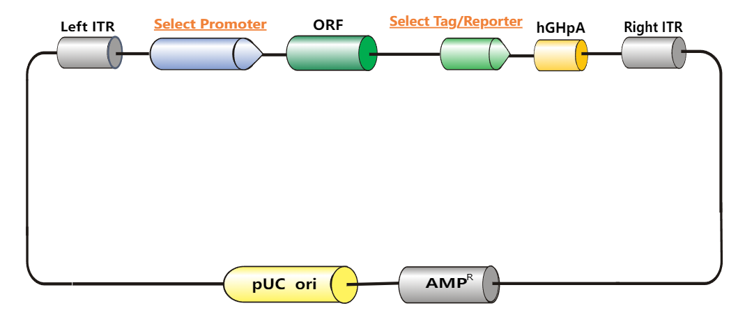
The destination vector carries your gene of interest (GOI) and two ITRs. You can choose from a variety of destination vectors carrying promoters including CMV, EF1a, CAG, CBh and other tissue-specific promoters, and reporter gene GFP for optimal gene expression and detection.(Learn more about our vector collection); the plasmid that carries the Rep and Cap genes from wild type AAV and the plasmid that carries adenovirus VA, E4 and E2A genes required for efficient AAV production are co-transfected with the destination vector for AAV packaging.
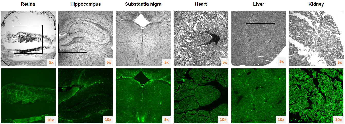
Figure 1. Purified eGFP AAV-9 Adeno-associated Viral Particles (Cat.No. AA016-100) was injected intravenously into adult mice at a dose of 1013 GC/kg (2-3 ×10¹¹ GC/ mouse), and tissue sections and fluorescence photography were performed after 2 weeks.
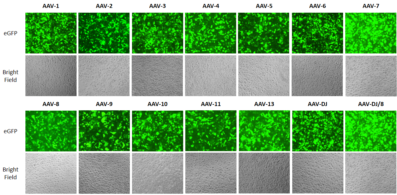
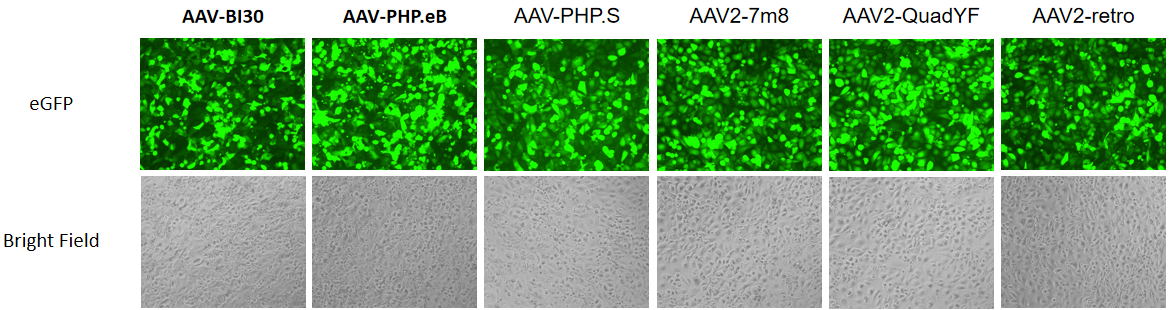
Figure 2. AAV of different serotypes carrying GFP were used to transduce the H1299 cell line as shown in the figure above. Fluorescent images (GFP) show that cells are transduced with AAV-GFP.
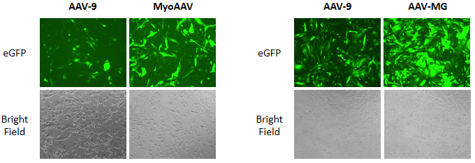
Figure 3. MyoAAV and AAV-MG indicated its tissue specificity in vitro infection. AAV-9, MyoAAV and AAV-MG carrying GFP were used to transduce the Rhabdomyosarcoma (RD) cell line(left) or Human microglia cell line (HmC3)(right).
AAV Serotype Choices
GeneCopoeia provides AAV for all the commonly-occurring AAV serotypes, including AAV1, AAV2, AAV3, AAV4, AAV5, AAV6, AAV7, AAV8, AAV9, AAV10, AAV11, AAV12, AAV13, AAV-DJ, AAV-DJ/8, MyoAAV, AAV-MG, AAV-PHP.eB and AAV-BI30, AAV-PHP.S, AAV2.7m8, AAV2-QuadYF, AAV2-retro. The intensity of gene expression and tissue tropism depends highly on the AAV serotype used in different tissues.
Different serotypes differ mainly in surface protein (capsid). Carefully choosing the serotype enables robust gene expression in specific tissues with minimal immune response. Refer to the table below to compare the essential differences between different serotypes.
AAV Serotypes Available
Serotype
Primary Target Tissues
Description
AAV-1
Muscle
Best for cardiac muscle, skeletal muscle, neuronal and glial tissue.
AAV-2
Muscle, Liver, Retina
Oldest and most commonly-used serotype. Best for neurons, muscle, liver, and brain.
AAV-3
Megakaryocytes
Best for megakaryocytes, muscle, liver, lung, and retina.
AAV-4
Retina
Best for neurons, muscle, brain, and retina.
AAV-5
Lung
Best for lung, neurons, synovial joint, retina.
AAV-6
Muscle, Lung
Best for lung, liver, and heart.
AAV-7
Muscle, Retina, Neurons
Best for muscle, neurons, and liver.
AAV-8
Liver
Best for muscle, brain, liver, retina, pancreas and adipose.
AAV-9
Various
Best for muscle, heart, liver, lung, and brain.
AAV-10
Pleura, CNS
Cloned from Cynomolgus, almost identical with AAVrh10 except for 12 amino acids in VP1. Best for lung, muscle, heart, CNS, and liver.
AAV-11 NEW!
Spinal nerves
Efficient retrograde targeting of projection neurons and enhances astrocyte-directed transduction.
AAV-12 NEW!
Salivary glands, Skeletal muscles
Does not depend on either heparan sulfate or sialic acid for transduction. Transduces salivary glands and skeletal muscles in vivo.
AAV-13 NEW!
CNS
Suitable for precise labeling, especially for targeting small nuclei in the brain.
AAV-DJ
Various
A mixture of 8 naturally-occurring serotypes. Efficiently transduces a wide variety of cell types in vitro.
AAV-DJ/8
Various
A variant of AAV-DJ with a mutation that permits infection of liver as well as other tissues in vivo.
MyoAAV NEW!
Muscle tissue
Best specificity for muscle tissue.
AAV-MG NEW!
Microglia
Best specificity for microglia in the brain.
AAV-PHP.eB NEW!
CNS
Cross the blood–brain barrier (BBB) and has enhanced CNS tropism.
AAV-BI30 NEW!
CNS
Specifically and efficiently transduces endothelial cells throughout the CNS.
AAV-PHP.S NEW!
PNS
Effective infection of peripheral nervous system.
AAV2.7m8 NEW!
Retina, inner ear
Effective infection of retinal cells, inner ear cochlear hair cells.
AAV2-QuadYF NEW!
Endothelial cell, retina
Effective infection of retinal cells, vascular endothelial cells.
AAV2-retro NEW!
Spinal nerves
Effective retrograde transduction of selective corticospinal tract neurons.
AAV6.2 NEW!
Retina, lung
Effective infection of retina, lung and the airway epithelium.
AAV9-insALAVPFR NEW!
Macrophage
Effective infection of non-CNS tissue-resident macrophages (TRMs).
We offer AAV of virtually all serotypes with titers starting from 5×10¹² GC/ml packaged with ORF cDNA, CRISPR/Cas9 or shRNA. Click the links below to search for your gene(s) of interest.
For custom AAV packaging service, please contact us at inquiry@genecopoeia.comto get a quote.
Choose from our collection of AAV vectors with different promoters and fluorescent markers for your gene expression. The AAV can be packaged with ORF cDNA clones up to 3 kb in length for long-term expression.
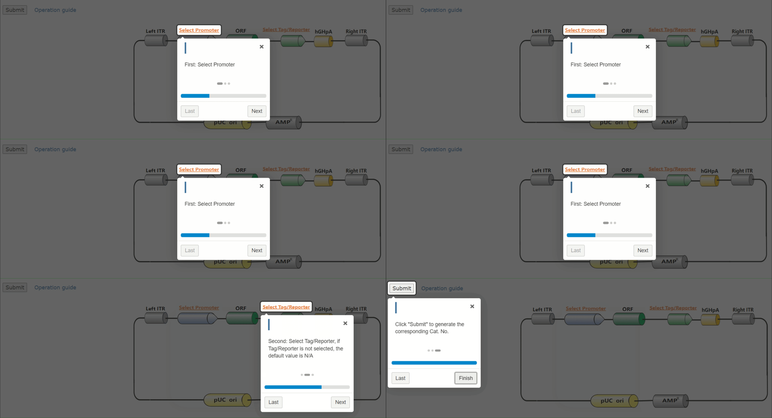
If none of our ORF expression-ready AAV vectors is suitable for your research, please use GeneCopoeia’s V-Architect tools which provides various promoters and tags to match your experimental design.
Transduction-ready AAV delivering shRNA of varying lengths (19 to 29 bases) that were designed using a proprietary algorithm to make shRNA expression constructs that have high knockdown efficiency with minimal off-target effect.
By default, all options for AAV serotype, gene of interest, and volume are listed in the table below. Use the drop-down menus to narrow down the number of options displayed.
EasyProbes™ Antifade Reagent is a liquid mountant applied directly to fluorescently labeled cell or tissue samples on microscope slides. It contains chemical components designed to protect fluorescent dyes from fading (photobleaching) during fluorescence microscopy experiments, and delivers this protection without significantly quenching the initial fluorescence signal. EasyProbes™ Antifade Reagent allows longer-term storage of the sample. It comes ready-to-use — just apply a drop to the sample, add a coverslip, cure, and image. EasyProbes™ Antifade Reagent is available at 10 ml bottle or 5×2 ml dropper bottle with or without DAPI nuclear stain.
Features
Protects dyes from fading during imaging
Ready-to-use liquid that cures for longer-term storage
Mounted samples are stable for months
Maintains fluorescence signal—little to no quenching



 Powered by Bioz