Select Page

sgRNA for CRISPR interference

Repress gene expression, without cutting the DNA

CRISPR interference (CRISPRi) offers significant advantages over traditional gene knockdown methods like RNAi, including highly specific, reversible silencing of genes at the DNA level without altering the genome sequence. It provides high-efficiency repression, low off-target effects, and high-throughput functionality for studying essential genes, noncoding RNAs, and complex pathways.
GeneCopoeia’s CRISPRi products and services provide a complete, powerful solution to your epigenetic editing needs.

sgRNA clones

Intended for use with dCas9-KRAB

sgRNA lentivirus particles

Transduction-ready particles, available in 3 purity levels

Vector choices

Vector Promoter CRISPR option Selection Marker / Reporter Gene Vector Type Vector Map
pCRISPR-AB04 U6 CRISPRi dCas9-KRAB Puro / mCherry Lentiviral get_info

 
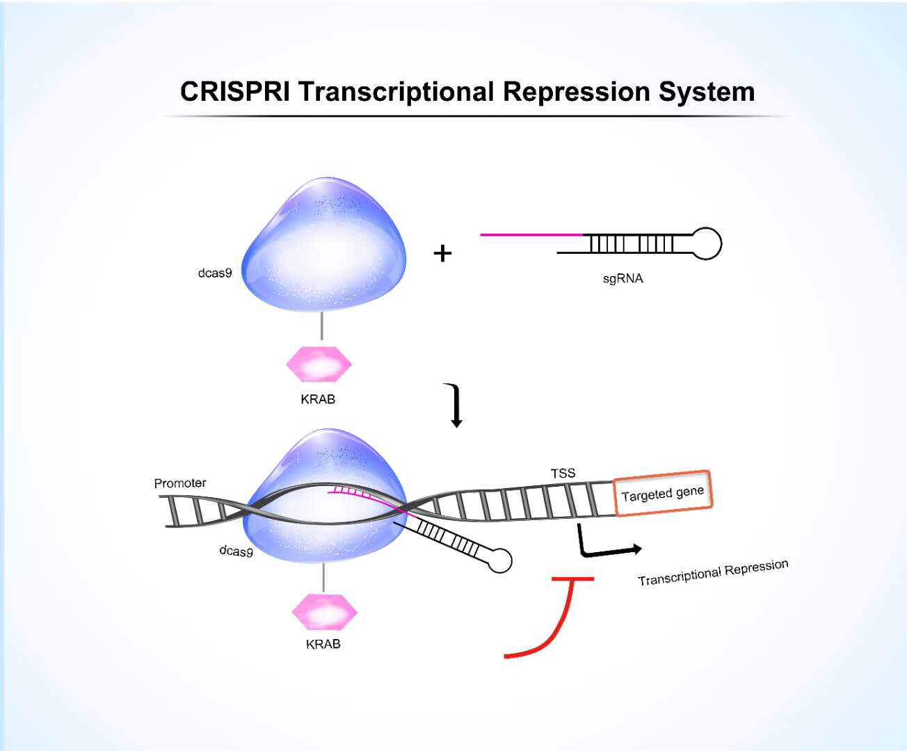
Technical overview The KRAB system is the most classic CRISPRi tool, achieved by fusing the Krüppel-associated box (KRAB) repression domain to nuclease-dead dCas9. Guided by an sgRNA, dCas9-KRAB specifically binds near the transcription start site (TSS) of the target gene. The KRAB domain then recruits the KAP1 co-repressor protein, initiating a cascade of epigenetic modification events. KRAB-CRISPRi is particularly useful for loss-of-function screening of essential genes, studying non-coding RNA regulation, and phenotypic analyses that mimic partial drug inhibition effects.

The CRISPRi system comprises 2 components
  • dCas9-KRAB fusion protein. Guided by the sgRNA, the dCas9 protein binds to the target DNA. Its fused KRAB domain then recruits epigenetic enzymes, such as the histone deacetylases HDAC1/2 and the histone methyltransferases, by binding to co-repressor proteins, such as KAP1. This process represses the transcriptional level of the target gene.
  • sgRNA

Application

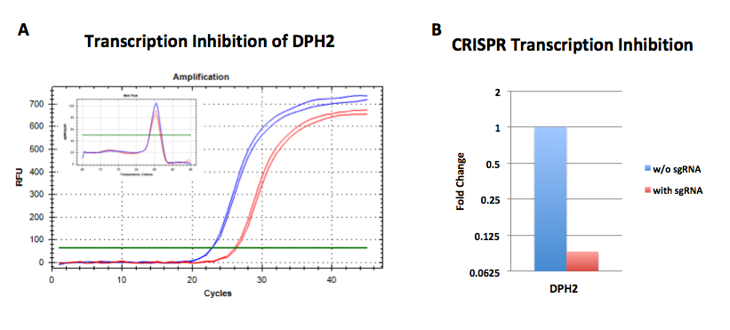
dCas9-KARB and sgRNA were co-overexpressed in HeLa cells. The transcriptional activity of DPH2 was assessed by qPCR. The amplification curves for cells transduced with sgRNA are shown in red, while those for cells without sgRNA transduction are shown in blue. A comparison of the transcriptional levels of the target gene with those of GAPDH demonstrated that the transcriptional levels of the target gene were significantly suppressed in HeLa cells.
sgRNA for CRISPR activation

Activate the transcription of any endogenous gene of interest, without cutting the DNA

CRISPR Activation (CRISPRa) is a powerful, precise gene-editing tool that upregulates specific endogenous genes without cutting the DNA. Key advantages include high specificity with minimal off-target effects, the ability to activate multiple genes simultaneously, and the versatility to regulate gene expression for therapeutic research or functional genomics without permanent genomic changes.
GeneCopoeia’s CRISPRa products and services provide a complete, powerful solution to your epigenetic editing needs.

sgRNA clones

Intended for use with dCas9 SAM

sgRNA lentivirus particles

Transduction-ready particles, available in 3 purity levels

Vector choices

Vector Promoter CRISPR option Selection Marker / Reporter Gene Vector Type Vector Map
pCRISPR-AG01 U6 CRISPRa dCas9 SAM Puro / eGFP Lentiviral get_info
pCRISPR-AG02 U6 CRISPRa dCas9 SAM Puro / eGFP Lentiviral get_info

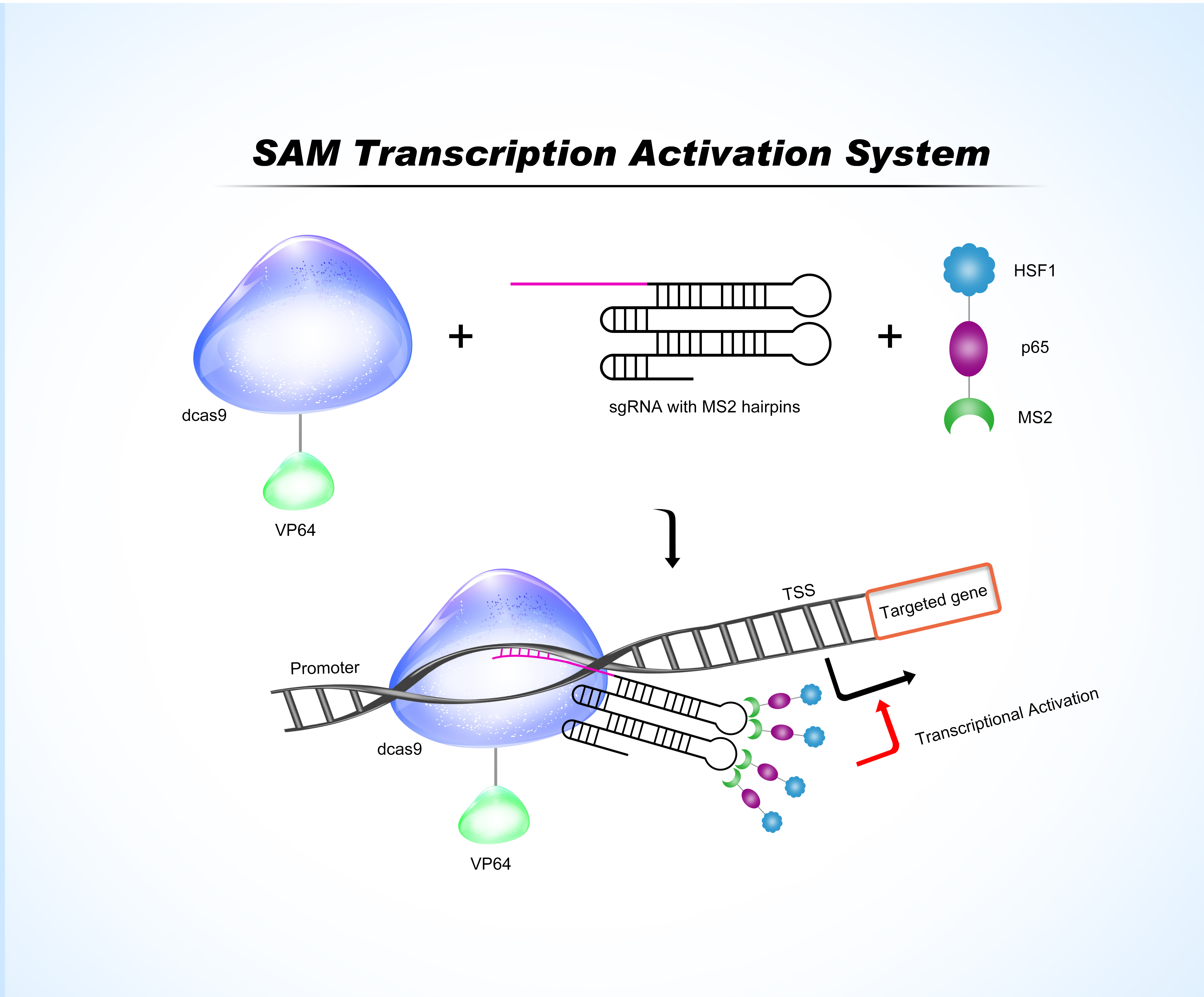
Technical overview The CRISPR (SAM) system (CRISPR-based Synergistic Activation Mediator) is designed to activate the transcription of any endogenous gene of interest. It is particularly useful for gain-of-function screening of endogenous genes, cellular reprogramming, and overexpression studies of key genes in disease models.

The system comprises 3 components that form a DNA-binding complex once transfected into the cells.
  • dCas9 fused to transcriptional activator VP64: VP64 acts as a strong transcriptional activator when recruited by a DNA-binding protein
  • sgRNA with an MS2 adaptor forming a characteristic stem-loop structure: The sgRNA-MS2 component forms a complex with dCas9 and directs it to the target DNA sequence next to the promoter region of the gene of interest
  • MCP-p65-HSF1 fusion protein (MPH complex): Once captured in the assembled complex at the gene promoter, P65 and HSF1 synergize with VP64 to robustly activate transcription of the downstream target gene by as much as a hundred-fold, depending on the gene

Application

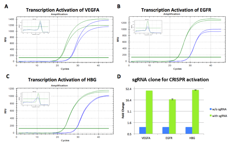
The MPH complex, dCas9-VP64, and sgRNA were co-overexpressed in HEK293 cells. The transcriptional activity of VEGFA (Figure A), EGFR (Figure B), and HBG (Figure C) was detected by qPCR. The cell proliferation curves for cells transduced with sgRNA are shown in green, while those for cells without sgRNA transduction are shown in blue. The transcriptional levels of the target genes were compared with those of GAPDH, indicating a significant increase in the transcriptional levels of the target genes in HEK293 cells.

sgRNA for CRISPRa and CRISPRi

CRISPR activation (CRISPRa) and interference (CRISPRi) are advanced genetic tools that utilize a catalytically “dead” Cas9 (dCas9) fused to effector proteins to modulate gene expression without cutting or altering the underlying DNA sequence. These technologies offer superior precision, reduced toxicity, and greater flexibility for regulating endogenous genes compared to traditional knockouts or cDNA overexpression.
GeneCopoeia’s CRISPR products and services provide a complete, powerful solution to your epigenetic editing needs.
 

Activate the transcription of any endogenous gene of interest, without cutting the DNA

Vector choices for expression clones and lentivirus particles

Vector Promoter CRISPR option Selection Marker / Reporter Gene Vector Type Vector Map
pCRISPR-AG01 U6 CRISPRa dCas9 SAM Puro / eGFP Lentiviral get_info
pCRISPR-AG02 U6 CRISPRa dCas9 SAM Puro / eGFP Lentiviral get_info
   

Repress gene expression, without cutting the DNA

Vector choices for expression clones and lentivirus particles

Vector Promoter CRISPR option Selection Marker / Reporter Gene Vector Type Vector Map
pCRISPR-AB04 U6 CRISPRi dCas9-KRAB Puro / mCherry Lentiviral get_info
 

OmicsArray™ Pre-made Peptide Array Services

Ready-to-Screen Panels for Infectious Disease, Autoimmunity, and Oncology

GeneCopoeia offers a diverse catalog of pre-designed peptide microarrays for full-service screening. These panels are designed by scientists to cover specific proteomes or immunodominant epitopes derived from the Immune Epitope Database (IEDB).  

Request the quote for the panel of your interest!

Autoimmune & Allergy Panels

High-density profiling of autoantibodies and allergen-specific IgE/IgG.

Cat.No. Panel Name Content & Description Documentation
PP001 Autoimmune Epitope Microarray 4,287 B-cell epitopes associated with ~50 autoimmune diseases (SLE, RA, MS).
PP201 Autoimmune Human Epitope Microarray Focused profiling of human-specific autoimmune targets (subset of PP001).
PP301 Lupus Microarray Specialized profiling for Systemic Lupus Erythematosus (SLE) autoantigens.
PP002 CNS Peptide Microarray Targets antigens related to Multiple Sclerosis and CNS disorders.
PP006 Allergen Epitope Microarray 4,022 B-cell epitopes from common environmental and food allergens.
PP031 Cyclic Citrullinated Peptide Microarray Screening for Rheumatoid Arthritis (RA) biomarkers using cyclic citrullinated peptides.
PP032 Human Cytokine Microarray Maps interactions with key human inflammatory cytokines and receptors.
PP033 Transglutaminase Peptide Microarray Targets tissue transglutaminase epitopes (Celiac Disease research).

OmicsArray™ Custom Peptide Array Service

High-Resolution Epitope Mapping & Discovery Services

Request a quote!

Overview

The OmicsArray™ Custom Peptide Array Service provides a fully tailored solution for researchers needing high-resolution epitope mapping, antibody profiling, and biomarker discovery.

Powered by PEPperPRINT® Laser Printing Technology, we synthesize custom peptide libraries directly on the chip surface. This “on-chip” synthesis allows for maximum flexibility and density, enabling us to print highly overlapping peptide libraries (e.g., shifting by just 1 amino acid) to map epitopes with single-residue precision.

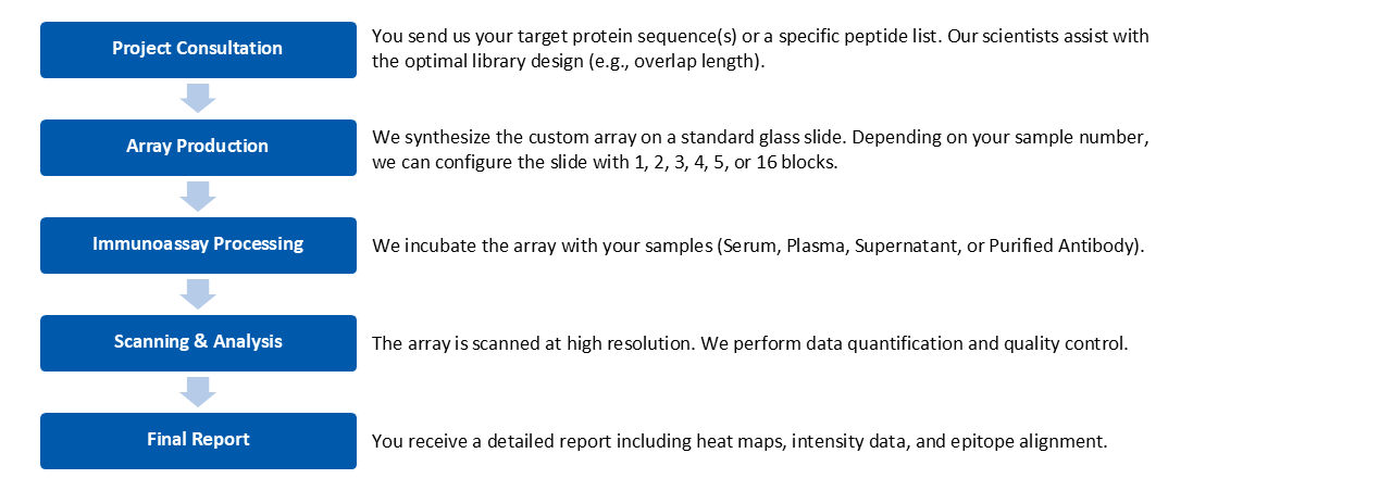
This is a Full-Service Solution: You provide the protein sequence or peptide list and your biological samples. Our team handles the entire workflow—library design, array synthesis, sample processing, scanning, and bioinformatics analysis.

Why Choose OmicsArray™ Custom Peptide Array Service?

  • Maximal Resolution: We typically use a “Shift of 1” design (15-mer peptides overlapping by 14 amino acids). This ensures that no binding motif is missed and allows for precise identification of essential binding residues.
  • Flexible Multiplexing (1 to 16 Blocks): Our standard slides can be printed as a single large array or divided into up to 16 individual blocks (sub-arrays). This allows you to screen multiple samples (up to 16) simultaneously on a single slide, maximizing cost-efficiency.
  • Cost-Effective Customization: Because we use digital laser printing, there are no expensive setup costs for masks or pre-synthesis of peptides.
  • Minimal Sample Required: We can generate high-quality data from as little as 20 µL of serum or plasma
 
Service Applications

Service Applications

We offer several custom design formats to suit your specific research goals.
  1. Linear Epitope Mapping                                                         The gold standard for defining antibody binding sites.
  • Method: We translate your target protein sequence into overlapping linear peptides.
  • Typical Design: 15 amino acid length with a 14 amino acid overlap (1 aa shift).
  • Goal: Pinpoint the exact linear sequence where your antibody or serum binds.
  1. Conformational Epitope Mapping (Cyclic Peptides)
  • mimic protein loops and 3D structures.*
  • Method: We synthesize peptides with cyclic constraints (e.g., disulfide bridges) directly on the array.
  • Typical Design: Loops of 7, 10, or 13 amino acids.
  • Goal: Identify antibodies that recognize 3D structural features rather than just linear sequences.
  1. Substitution Scans (“Alanine Scanning”)                                 Identify critical residues for binding affinity.
  • Method: Systematically substitute every amino acid in a known wild-type epitope with Alanine (or other amino acids).
  • Goal: Determine which specific amino acids are essential for the antibody-antigen interaction.
  1. Discovery Screening                                                                   For unbiased discovery.
  • Method: Screen randomized libraries or combinatorial lists.
  • Goal: Discover novel mimotopes or signature biomarkers from complex samples.